44 label the structures of a skeletal muscle fiber.
Art-labeling Activity: The Structure of a Skeletal Muscle Fiber - Quizlet Start studying Art-labeling Activity: The Structure of a Skeletal Muscle Fiber. Learn vocabulary, terms, and more with flashcards, games, and other study tools. Skeletal Muscle Fiber Definition and Anatomy - Study.com The three types of skeletal muscle fiber types are type 1, type 2A, and type 2B. Type 1 fibers are slow-twitch muscle fibers, type 2A is fast-twitch intermediate fibers, and type 2B are fast ...
To label: The given structure in the diagram of a skeletal muscle fiber ... Science Biology Visual Essentials of Anatomy &Physiology To label: The given structure in the diagram of a skeletal muscle fiber. Introduction: Skeletal muscles are made up of skeletal muscle fibers and connective tissues. The important function of skeletal muscle is to allow the intentional or voluntary movement of the body parts.
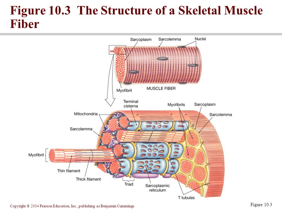
Label the structures of a skeletal muscle fiber.
Skeletal Muscle Fiber - GetBodySmart Skeletal muscles are a type of striated muscle. They are attached to the bones of the skeleton by tendons. Skeletal muscle fibers have a striated (striped) appearance because they are made up of smaller units called sarcomeres that run parallel to each other. Skeletal Muscle Fiber Labeling - Printable About this Worksheet. This is a free printable worksheet in PDF format and holds a printable version of the quiz Skeletal Muscle Fiber Labeling. By printing out this quiz and taking it with pen and paper creates for a good variation to only playing it online. Label structure of skeletal muscle Diagram | Quizlet Label structure of skeletal muscle 4.0 5 Reviews How do you want to study today? Learn Focus your studying with a path Test Take a practice test Match Get faster at matching terms + − Created by danielaaaa04 Terms in this set (8) myofibrils ... sarcoplasmis reticulum ... sarcolemma ... epimysium ... perimysium ... endomysium ... fascicle ...
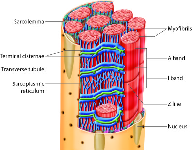
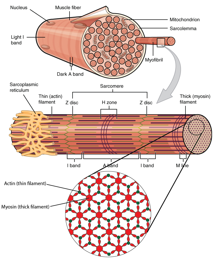
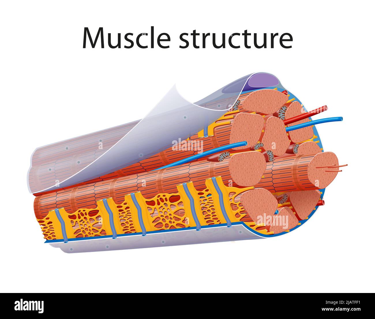
Label the structures of a skeletal muscle fiber.. Skeletal Muscle - Anatomy & Physiology - University of Hawaiʻi Muscles attach to bones directly or through tendons or aponeuroses. Skeletal muscles maintain posture, stabilize bones and joints, control internal movement, and generate heat. Skeletal muscle fibers are long, multinucleated cells. The membrane of the cell is the sarcolemma; the cytoplasm of the cell is the sarcoplasm. Chapter 9 Homework Flashcards | Quizlet Review some terms associated with a skeletal muscle fiber (also called a muscle cell) and the sarcomere. Drag and drop each term to the best description of that term. Review another set of terms associated with a skeletal muscle fiber/cell and the sarcomere. Internal Anatomy of Skeletal Muscle Fibers | GetBodySmart Learn more about muscle anatomy with these efficient, interactive exam-style quizzes. Learn anatomy faster and. remember everything you learn. Start Now. <. General Anatomy of Skeletal Muscle Fibers. >. Skeletal Muscle Fiber Structure and Function - Open Textbooks for Hong Kong Within each muscle fiber are myofibrils, long cylindrical structures that lie parallel to the muscle fiber. Myofibrils run the entire length of the muscle fiber. They attach to the plasma membrane, called the sarcolemma, at their ends, so that as myofibrils shorten, the entire muscle cell contracts ( Figure 16.18 ).
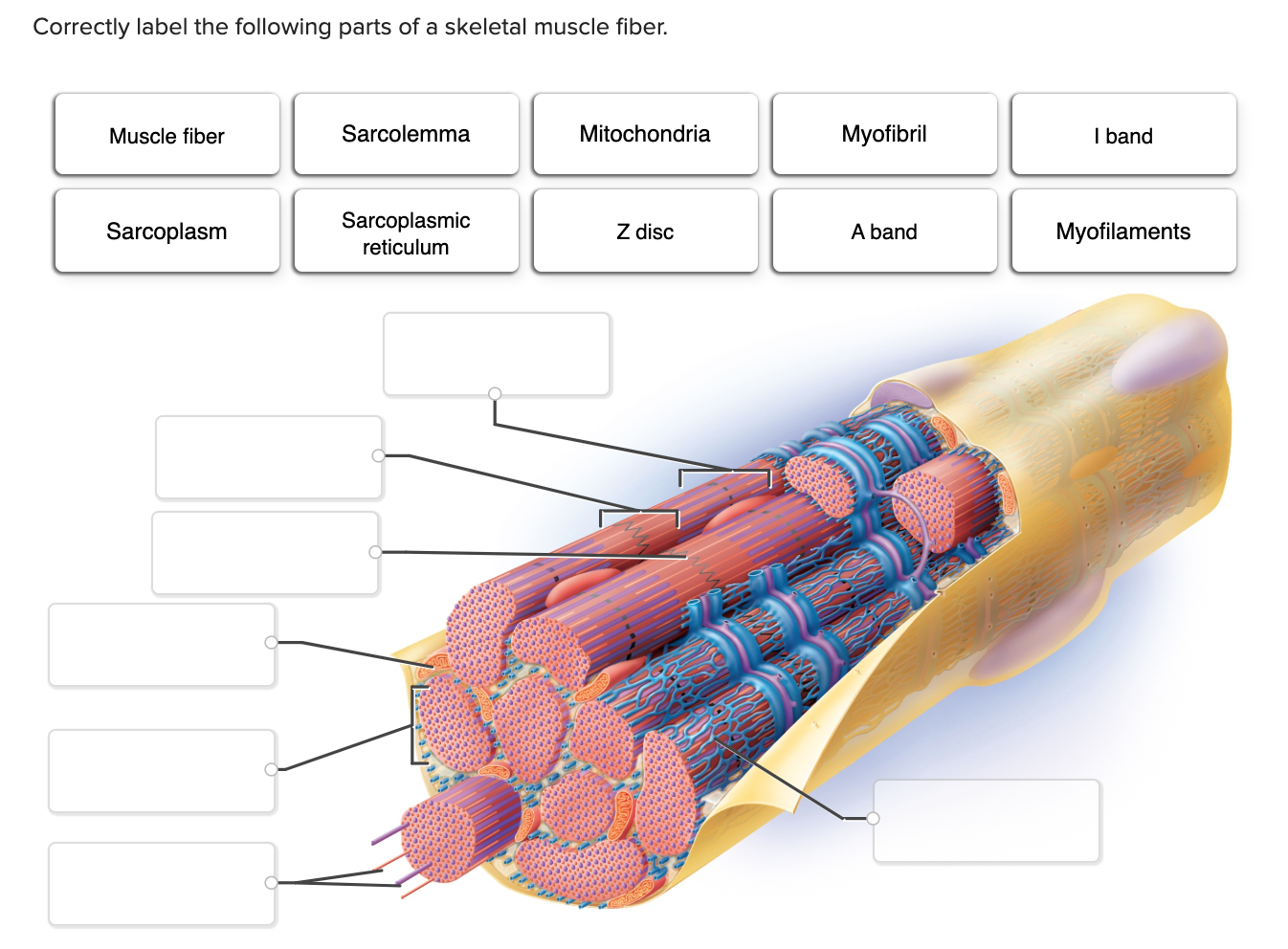
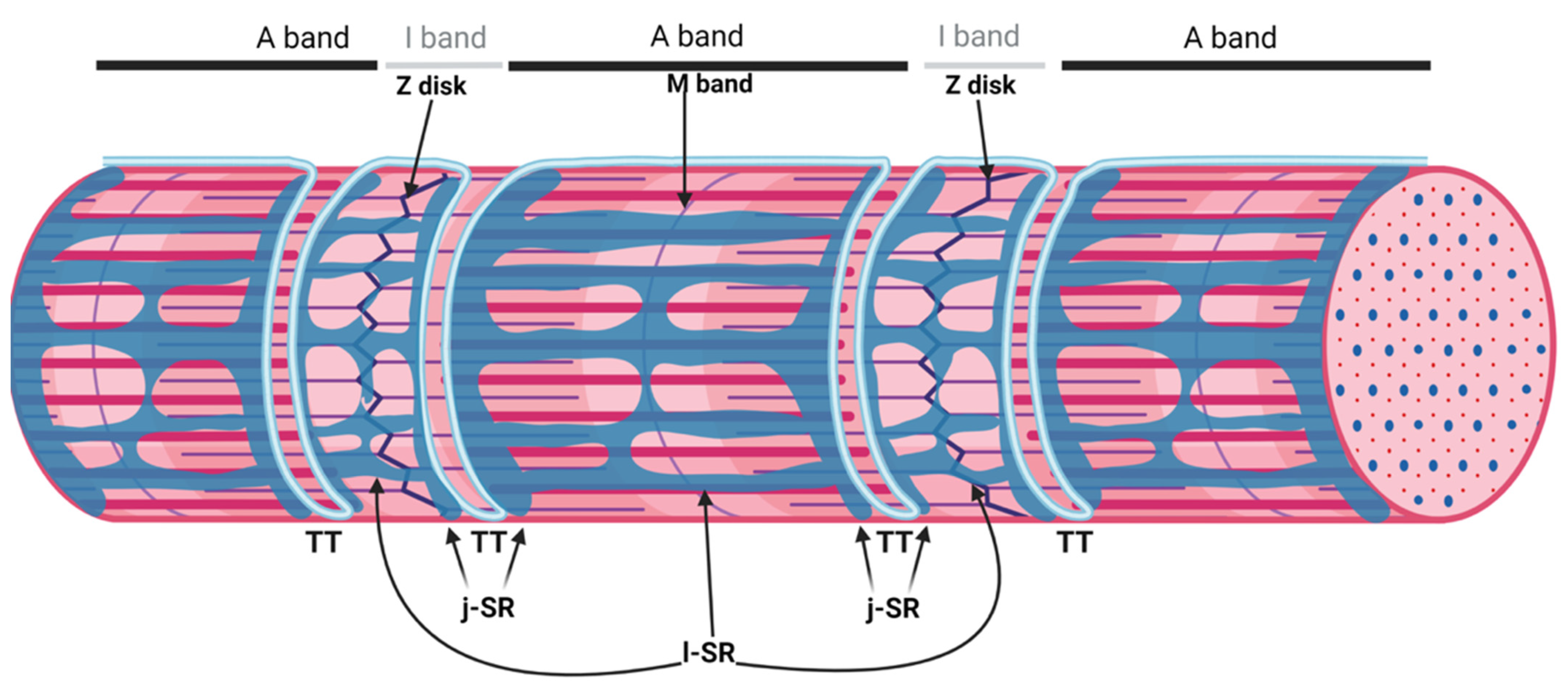
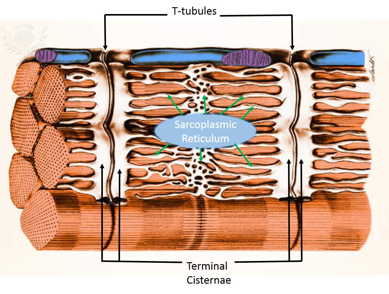
Correctly Label The Following Parts Of A Skeletal Muscle Fiber A skeletal muscle fiber is surrounded by a plasma membrane, which contains the sarcoplasm of the muscle cell. It is composed of many fibrils. These fibrils give the muscle fiber its striated appearance. The myofilaments are arranged sequentially in the sarcomere, which gives the fiber its striated look. Label the image of a skeletal muscle fiber below. Actin Muscle fascicle ... 1. Describe the structure of a muscle, including the following terms: myosin, actin, sarcomere, myofibril, fiber, bundle. Identify the muscle structure indicated with each number and match it to the appropriate label in... Identify the muscle structure indicated with each number and match it to the appropriate label in the table. Muscle Fibers: Anatomy, Function, and More - Healthline Each one of your skeletal muscles is made up of hundreds to thousands of muscle fibers that are tightly wrapped together by connective tissue. Each muscle fiber contains smaller units made up of... Structures of the Skeletal Muscle Fiber Flashcards | Quizlet muscle's plasma membrane, wraps fibers of muscle T-tubule along middle of each terminal cisterna, inward extension of sarcolemma terminal cisterna enlarged areas of the sarcoplasmic reticulum surrounding the transverse tubules sarcoplasmic reticulum modified endoplasmic reticulum that wraps around myofibrils; part of sarcoplasm myofibrils
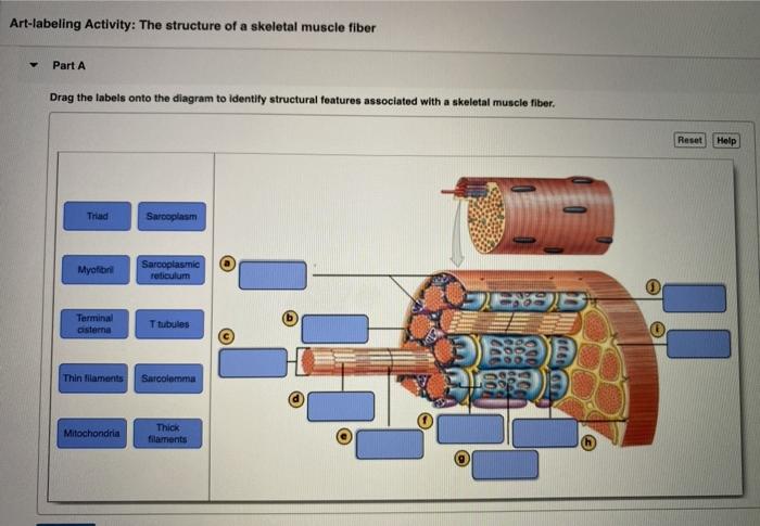
Solved Hel Label the structures of a skeletal muscle fiber ... - Chegg Science. Anatomy and Physiology. Anatomy and Physiology questions and answers. Hel Label the structures of a skeletal muscle fiber. 4 0.1 points eBook Sarcoplasmic reticulum Nucleus Myofibril Openings into T tubules Sarcolemma Mc Graw Hill < Prey 4 of 20 !!! AnatomyZone - Your Guide to Human Anatomy AnatomyZone is the leading resource for simple and concise 3D anatomy tutorials, with over 200 videos and a new range of interactive 3D anatomy models. SKELETAL MUSCLE ORGANIZATION - Brigham Young University-Idaho Skeletal Muscle Cells-Gross and Microscopic Structure. Each skeletal muscle cell, also called a muscle fiber, develops as many embryonic myocytes fused into one long, multi-nucleated skeletal muscle cell. These muscle fibers are bound together into bundles, or fascicles, and are supplied with a rich network of blood vessels and nerves. The ... Fibers of the skeletal muscle | Anatomy snippets The percentage of each muscle fiber present in an individual is determined by three factors: genetics, hormone levels within the blood, and the level of training undertaken. The skeletal muscle is one of 14 microanatomy models on the Complete Anatomy platform. Experience the minute detail of the human body in stunning 3D.
Muscles Labeling
Skeletal Muscle Histology Slide Identification and Labeled Diagram ... Please try to find out these structures from the skeletal muscle slide labeled images. #1. Longitudinal section of skeletal muscle #2. Cross-section of skeletal muscle #3. Skeletal muscle fibers of the longitudinal section #3. The nucleus of skeletal muscle fibers in longitudinal and cross-section #4. Cross striations of skeletal muscles #5.

Identify the structures of skeletal muscle. - Brainly.com
Skeletal Muscle Fiber Labeling Quiz - purposegames.com This is an online quiz called Skeletal Muscle Fiber Labeling There is a printable worksheet available for download here so you can take the quiz with pen and paper. Your Skills & Rank Total Points 0 Get started! Today's Rank -- 0 Today 's Points One of us! Game Points 15 You need to get 100% to score the 15 points available Actions
Muscular Levels of Organization | Anatomy and Physiology I ...
Nervous System: Explore the Nerves with Interactive Anatomy ... Nov 02, 2020 · Efferent neurons (also called motor neurons) carry signals from the gray matter of the CNS through the nerves of the peripheral nervous system to effector cells. The effector may be smooth, cardiac, or skeletal muscle tissue or glandular tissue. The effector then releases a hormone or moves a part of the body to respond to the stimulus.
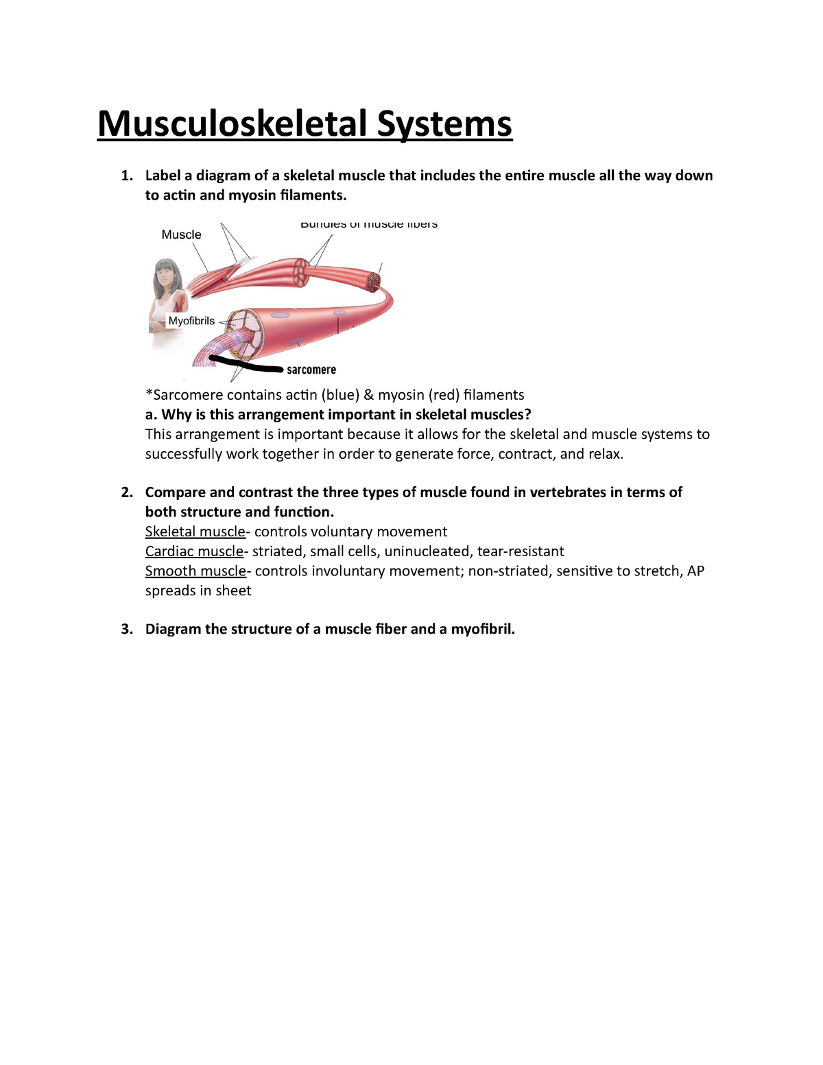
Musculoskeletal Systems - Label a diagram of a skeletal ...
Phosphoproteomics of three exercise modalities identifies ... Jul 25, 2022 · One of these core phosphosites was S67 on the uncharacterized protein C18ORF25, which we validated as an AMPK substrate. Mice lacking C18ORF25 have reduced skeletal muscle fiber size, exercise capacity, and muscle contractile function, and this was associated with reduced phosphorylation of contractile and Ca 2+ handling proteins. Expression of ...
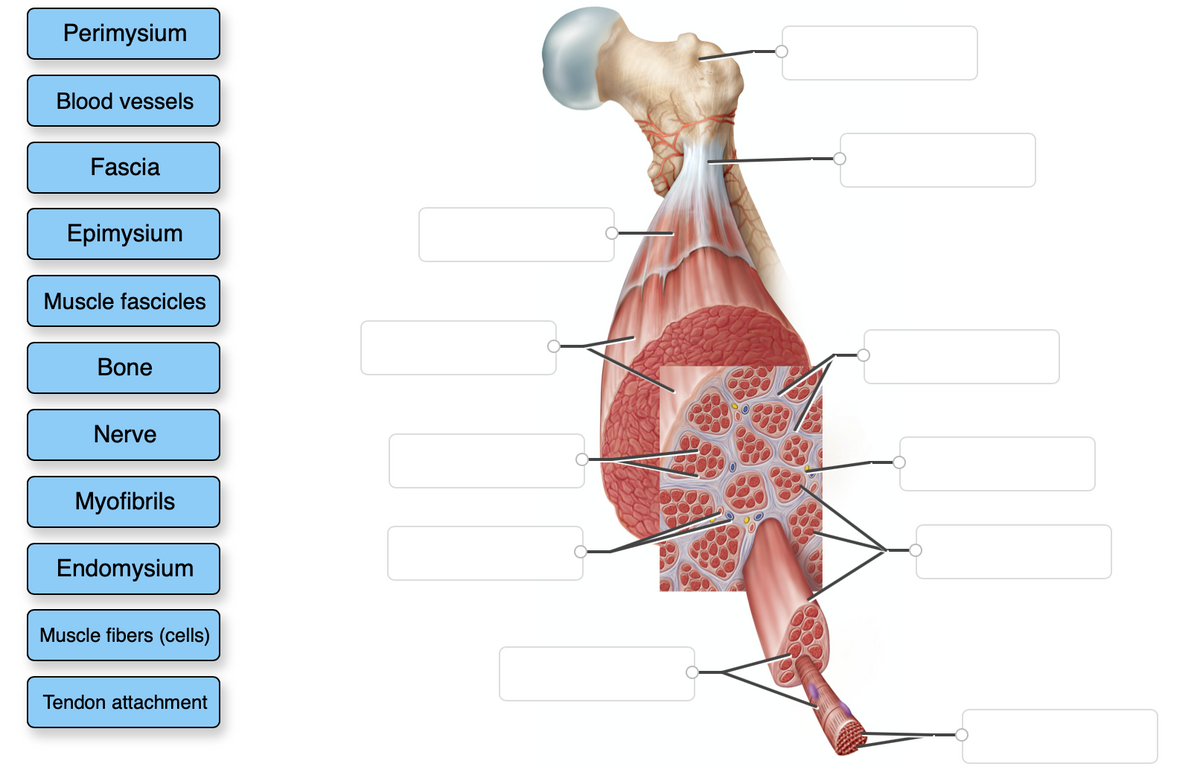
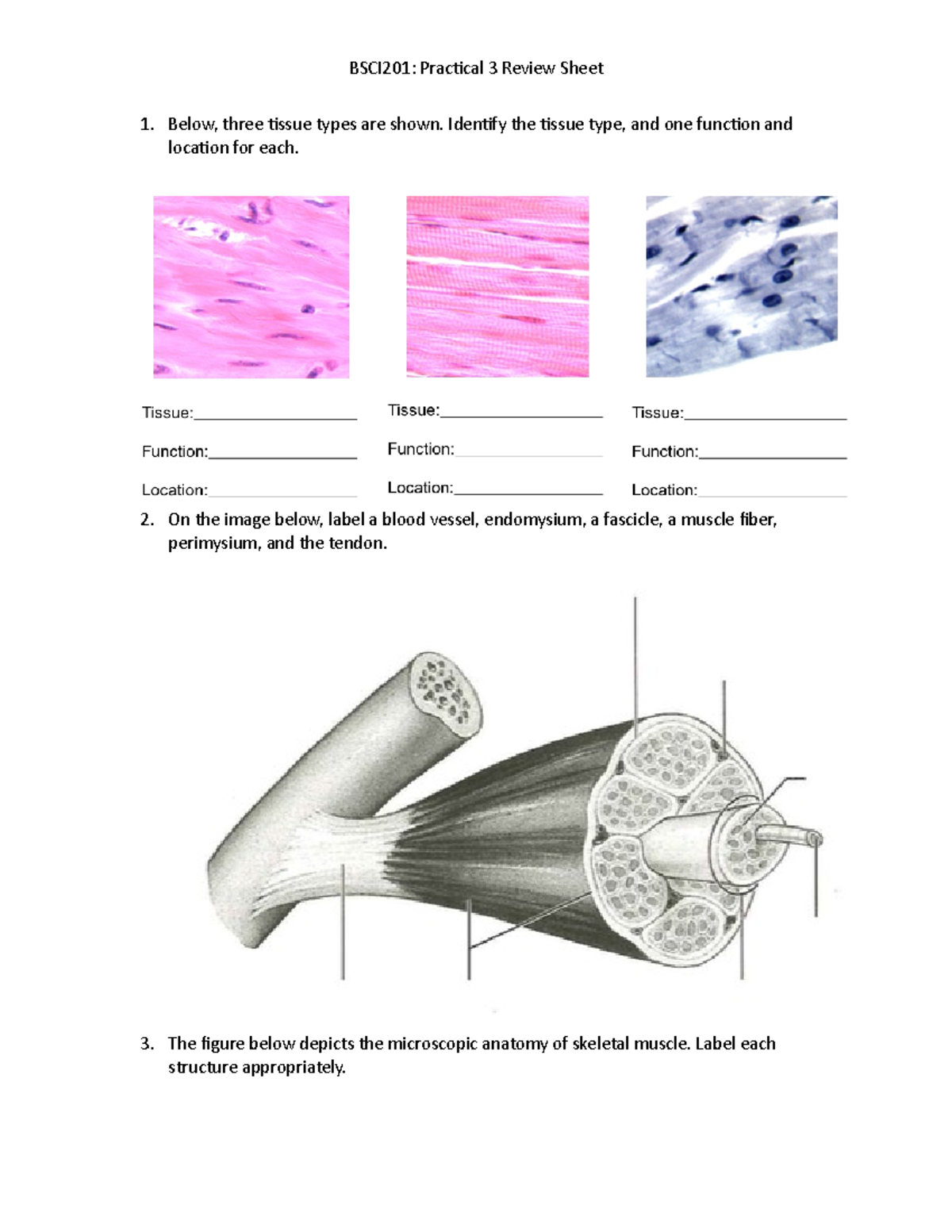
Answered: Perimysium Blood vessels Fascia… | bartleby
structure of a skeletal muscle fiber labeled Diagram | Quizlet structure of a skeletal muscle fiber labeled STUDY Terms in this set (...) myosin (thick filaments) actin (thin filaments) transverse tubule sarcoplasmic reticulum triad myofibrils sarcolemma nucleus transvertubule nucleus Muscle fiber14 terms namba8hcc Anatomy chapter 9 skeletal muscle fibers14 terms LeeFitz Muscle cell = Muscle fiber11 terms
Solved Correctly label the following parts of a skeletal ...
Skeletal Muscle | Anatomy and Physiology I - Lumen Learning A skeletal muscle fiber is surrounded by a plasma membrane called the sarcolemma, which contains sarcoplasm, the cytoplasm of muscle cells. A muscle fiber is composed of many fibrils, which give the cell its striated appearance. The Sarcomere
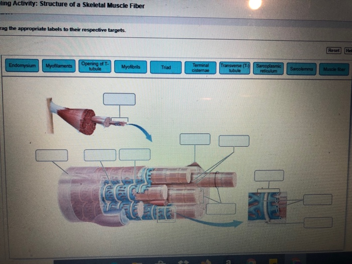
Solved -ling Activity: Structure of a Skeletal Muscle Fiber ...
General Anatomy of Skeletal Muscle Fibers | GetBodySmart Internal Anatomy of Skeletal Muscle Fibers. An interactive quiz about the internal anatomy of skeletal muscle fibers, featuring illustrations-based multiple choice questions. Skeletal Muscle Fiber Location and Arrangement > Internal Anatomy of Skeletal Muscle Fibers. Subject Areas. Skeletal System; Muscular System;

Muscle Fiber (3B) - Pierce College - Anatomy
Septin7 is indispensable for proper skeletal muscle ... - eLife Aug 05, 2022 · Septin7 is described as a novel component of skeletal muscle cytoskeleton with essential roles in muscle development and the proper organization of the myofilaments and the mitochondrial network while its absence leeds to reduced force and skeletal deformities.

How Muscle Structure and Composition Influence Meat and Flesh ...
Solved Muscle Cell Label the structures of a skeletal muscle - Chegg 100% (17 ratings) 1) Sarcolemma 2) myofib … View the full answer Transcribed image text: Muscle Cell Label the structures of a skeletal muscle fiber. Nucleus Myofibril Sarcolemma Sarcoplasmic reticulum Openings into T tubules < Prev 3 of 15 !!! Next > Thinkinys - How to write a boty The Good Cre. Dob C ommunicatio pdf Communication.pdf

The structure and internal organization of a skeletal muscle ...
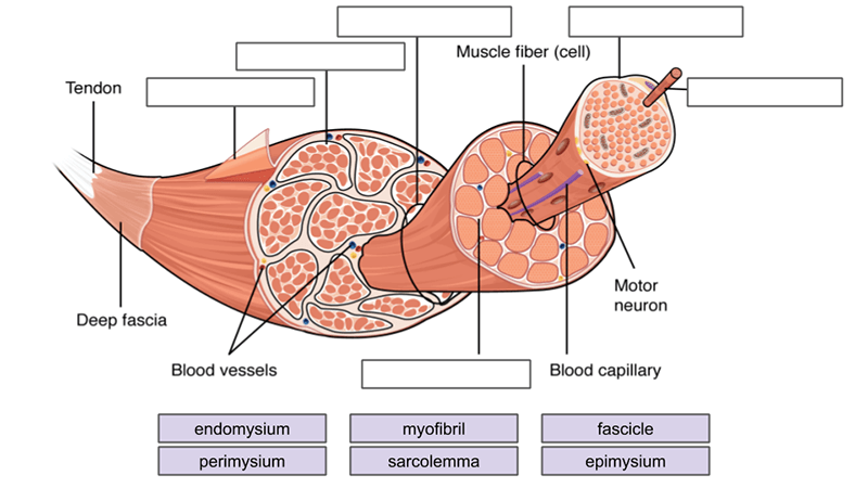
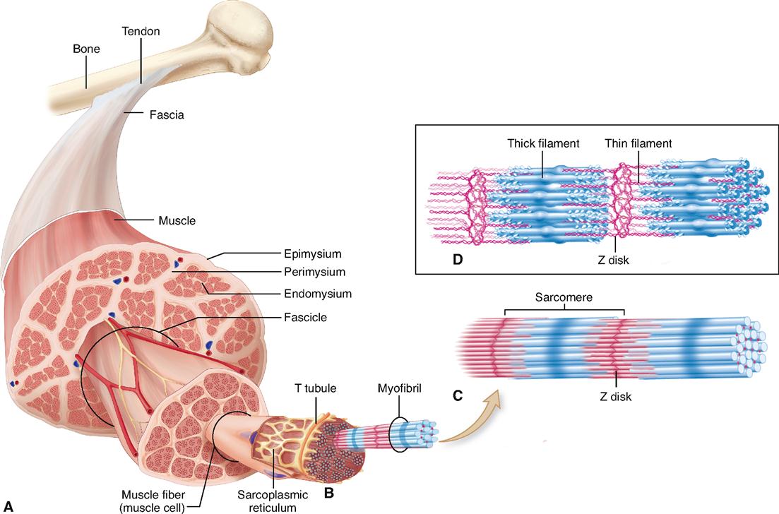
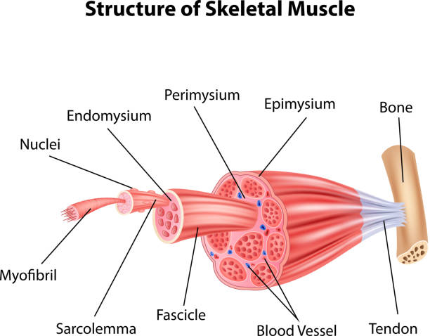
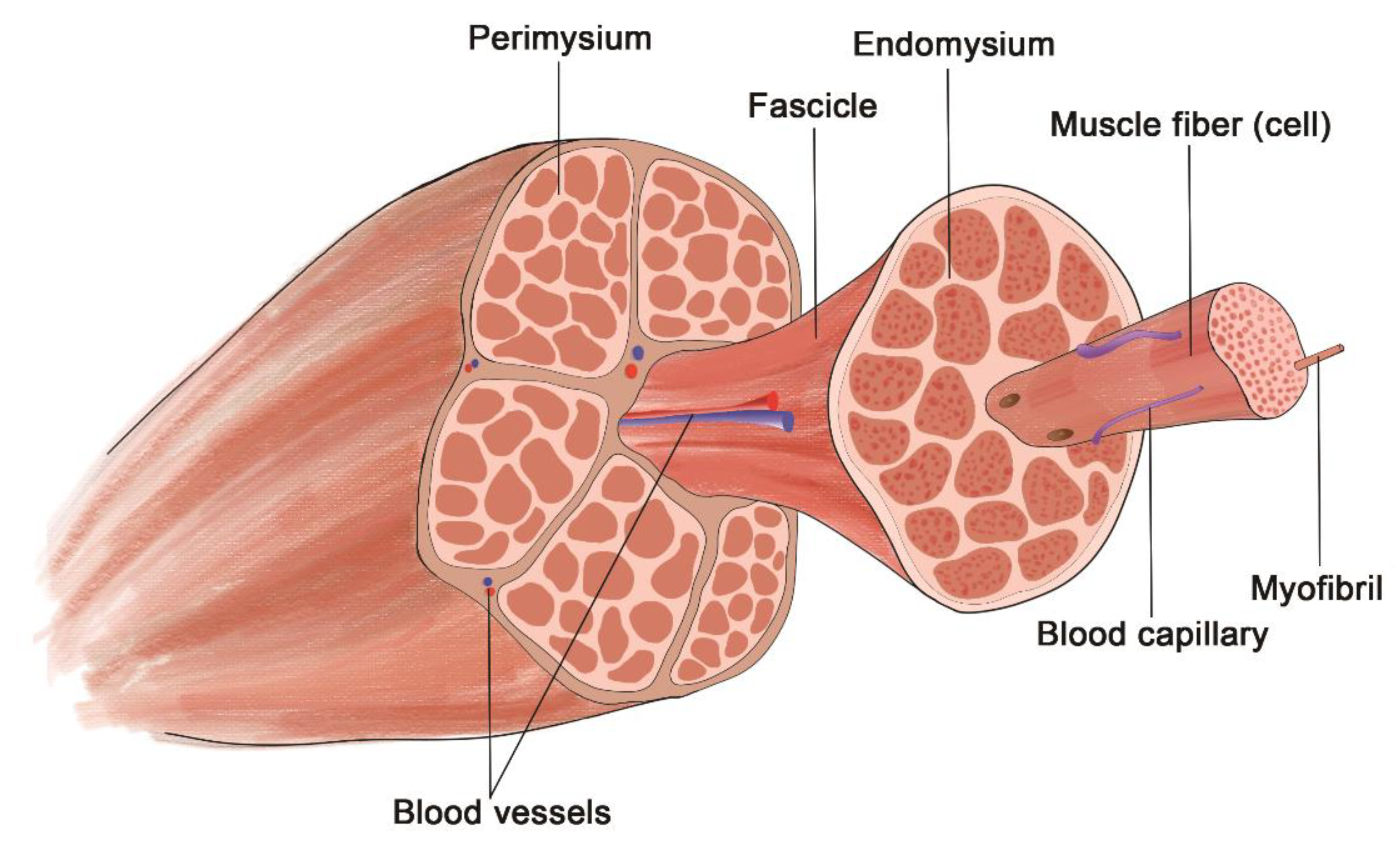
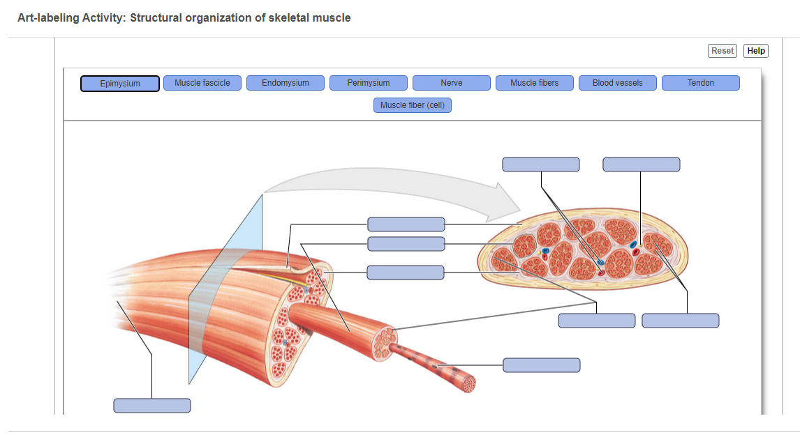
Draw the basic structure of skeletal muscle. Label the following ... SECTION 1: Structure of a Skeletal Muscle A. Layers of connective tissue separate a muscle into compartments. The layer called endomy- sium surrounds each muscle cell. Muscle cells are then bundled into fascicles, which are sur- rounded by perimysium. The entire muscle is composed of many fascicles, and is surround- ed by epimysium.
Place the following steps in order of how a muscle contracts ...
Phosphoproteomics of three exercise modalities identifies ... Jul 25, 2022 · Exercise induces signaling networks to improve muscle function and confer health benefits. To identify divergent and common signaling networks during and after different exercise modalities, we performed a phosphoproteomic analysis of human skeletal muscle from a cross-over intervention of endurance, sprint, and resistance exercise.

Solved ge Home Topic Application Technolog.. Seved 19 Label ...
Structure of Skeletal Muscle | SEER Training Each organ or muscle consists of skeletal muscle tissue, connective tissue, nerve tissue, and blood or vascular tissue. Skeletal muscles vary considerably in size, shape, and arrangement of fibers. They range from extremely tiny strands such as the stapedium muscle of the middle ear to large masses such as the muscles of the thigh.

Skeletal Muscle | Anatomy and Physiology I | | Course Hero
Skeletal Muscle Fiber Labeled Muscle skeletal transverse labeled tissue section. Skeletal Muscle Fiber Labeled. SIU SOM Histology SSB we have 9 Pics about SIU SOM Histology SSB like Slide 34 - Skeletal Muscle - YouTube, Microscopic structure of skeletal muscle by Dr. S. N. Singh and also Microscopic structure of skeletal muscle by Dr. S. N. Singh. Here you go:

Schematic drawing illustrating the concept of the proposed ...
Week 6: Muscle Physiology Flashcards & Practice Test | Quizlet Sodium channel : a type of voltage-gated ion channel located on the sarcolemma of the muscle fiber. Acetylcholine : neurotransmitter that stimulates skeletal muscle contraction. Acetylcholinesterase : enzyme located in the synaptic cleft that breaks down acetylcholine. Synaptic cleft : the space between the axon terminal and junctional folds.
SKELETAL MUSCLE ORGANIZATION
Skeletal Muscle Fiber Labeling Flashcards | Quizlet Start studying Skeletal Muscle Fiber Labeling. Learn vocabulary, terms, and more with flashcards, games, and other study tools. Home. Subjects. Explanations. Create. Study sets, textbooks, questions ... Laboratory Manual for Hole's Essentials of Human Anatomy & Physiology 12th Edition Terry R. Martin. 1,633 explanations. Sets found in the same ...
Chapter 10
Skeletal Muscle Tissue Anatomy and Structure - Registered Nurse RN Each skeletal muscle is considered an organ, and it's made up of connective tissue layers, muscle fibers, blood vessels, and nerves. Skeletal muscles attach to the bones through tendons or through a direct attachment. As you look at this muscle diagram, you'll notice an outer layer of connective tissue called epimysium.
Biomolecules | Free Full-Text | The Sarcoplasmic Reticulum of ...
Skeletal muscle tissue: Histology | Kenhub Special terms are used to describe structures associated with skeletal muscle tissue. Muscle tissue terms often begin with myo-, mys-, or sarco-. The cytoplasm of a muscle cells is referred to as sarcoplasm.The plasma membrane is called the sarcolemma and the endoplasmic reticulum is called the sarcoplasmic reticulum.A muscle fiber may also be referred to as a myofiber.

Study Guide Flashcards | Quizlet
Label structure of skeletal muscle Diagram | Quizlet Label structure of skeletal muscle 4.0 5 Reviews How do you want to study today? Learn Focus your studying with a path Test Take a practice test Match Get faster at matching terms + − Created by danielaaaa04 Terms in this set (8) myofibrils ... sarcoplasmis reticulum ... sarcolemma ... epimysium ... perimysium ... endomysium ... fascicle ...
Muscle Tissue. - ppt download
Skeletal Muscle Fiber Labeling - Printable About this Worksheet. This is a free printable worksheet in PDF format and holds a printable version of the quiz Skeletal Muscle Fiber Labeling. By printing out this quiz and taking it with pen and paper creates for a good variation to only playing it online.
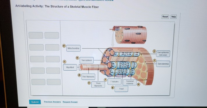
Solved Art-labeling Activity: The Structure of a Skeletal ...
Skeletal Muscle Fiber - GetBodySmart Skeletal muscles are a type of striated muscle. They are attached to the bones of the skeleton by tendons. Skeletal muscle fibers have a striated (striped) appearance because they are made up of smaller units called sarcomeres that run parallel to each other.

Structure of a Skeletal Muscle Fiber Quiz
Solved] Label all the parts of the follow diagrams. | Course Hero

1,998 Skeletal Muscle Stock Photos, Pictures & Royalty-Free ...
Anatomy Review: Skeletal Muscle Tissue

Label structure of skeletal muscle Diagram | Quizlet
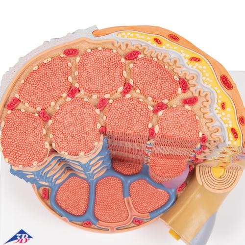
3B MICROanatomy™ Human Muscle Fiber Model, 10,000 times magnified - 3B Smart Anatomy
Physiology of the Muscular System | Basicmedical Key

Week 6: Muscle Physiology Flashcards | Quizlet
SKELETAL MUSCLE ORGANIZATION
3,366 Muscle Cells Stock Photos, Pictures & Royalty-Free ...

Biochemical and structural basis of the passive mechanical ...
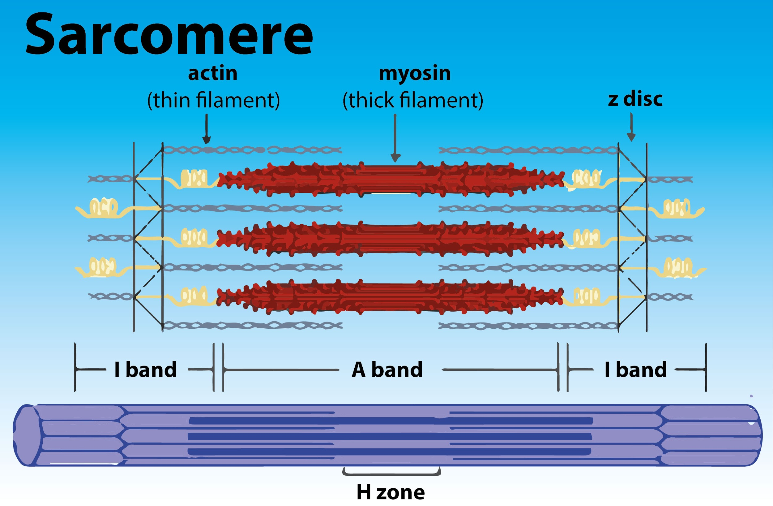
Draw the diagram of a sarcomere of skeletal muscle class 11 ...
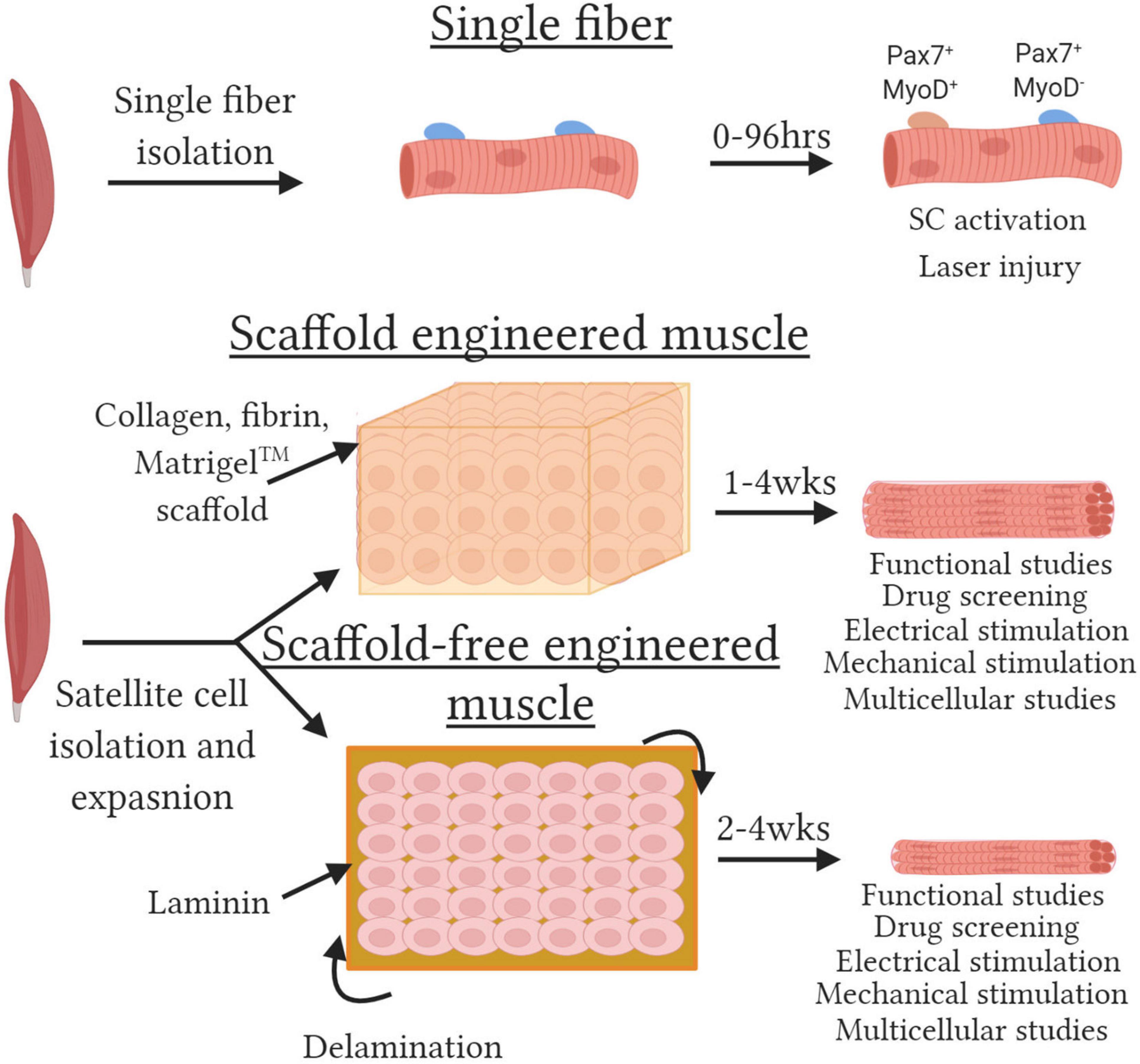
Frontiers | Tissue-Engineered Skeletal Muscle Models to Study ...

cumulative topic 6: microanatomy of myofiber | Body muscle ...
Polymers | Free Full-Text | A Review of Recent Advances in ...
Review of Muscles - 1. Below, three tissue types are shown ...

10.2 Skeletal Muscle – Anatomy & Physiology

Solved Muscle Cell Label the structures of a skeletal muscle ...

Ultrastructure of a skeletal muscle fiber. (a) Arrangement of ...
Skeletal muscle hi-res stock photography and images - Alamy
Solved Art-labeling Activity: The Structure of a Sarcomere ...

Lesson Explainer: Structure of Muscles | Nagwa
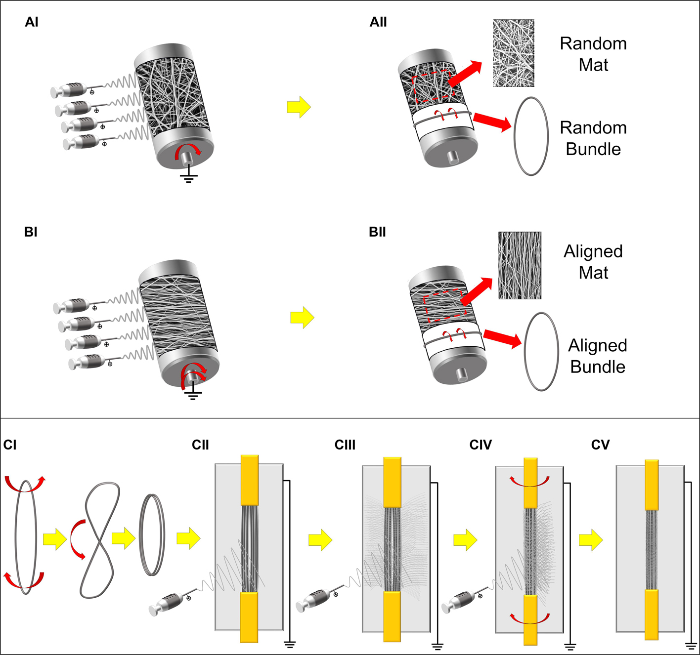
Frontiers | Biomimetic Hierarchically Arranged Nanofibrous ...
Answered: Art-labeling Activity: Structural… | bartleby
Post a Comment for "44 label the structures of a skeletal muscle fiber."