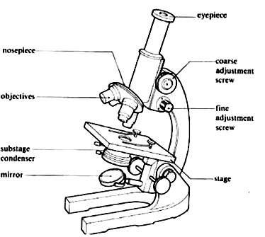
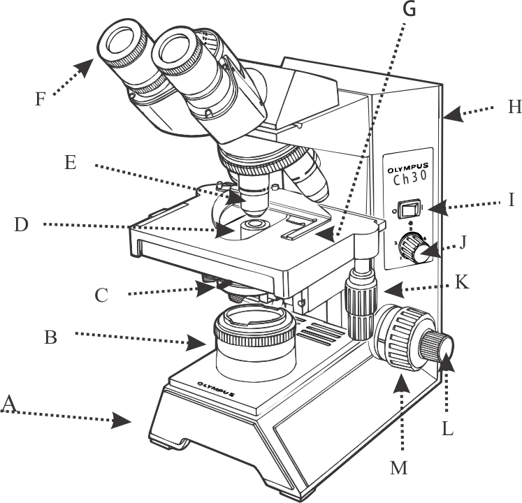
45 microscope drawing and label
Metaphase - Genome.gov During metaphase, the nucleus dissolves and the cell's chromosomes condense and move together, aligning in the center of the dividing cell. At this stage, the chromosomes are distinguishable when viewed through a microscope. Metaphase chromosomes are used in karyotyping, a laboratory technique for identifying chromosomal abnormalities. Narration Single cell atlas of spinal cord injury in mice reveals a pro ... After bilateral spinocerebellar labeling and 3 weeks post thoracic injury, we found that 65.2% (±4.3) of Sox11-expressing neurons, 41.8% (±1.5) of Sprr1a-expressing neurons and 38.0% (±5.6 SEM ...
Graphene - Wikipedia Graphene (/ ˈ ɡ r æ f iː n /) is an allotrope of carbon consisting of a single layer of atoms arranged in a two-dimensional honeycomb lattice nanostructure. The name is derived from "graphite" and the suffix -ene, reflecting the fact that the graphite allotrope of carbon contains numerous double bonds.. Each atom in a graphene sheet is connected to its three nearest neighbors by a strong ...

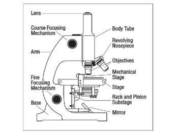
Microscope drawing and label
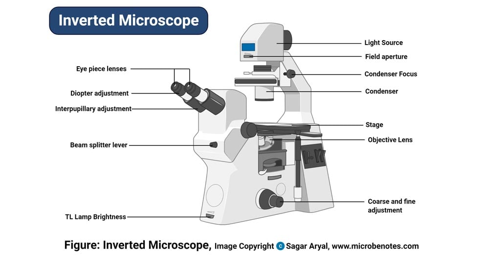
Spiny chondrichthyan from the lower Silurian of South China Circles opposite the taxon labels show the presence of these characters at terminal nodes. ... (V27437.3 and V27437.5-7) were imaged with a GXM XTL3101 stereo microscope at the School of ... ECLIPSE Ti2 Series | Inverted Microscopes | Nikon Microscope Products ... The ECLIPSE Ti2 inverted microscope delivers an unparalleled 25mm field of view (FOV) that revolutionizes the way you see. With this incredible FOV, the Ti2 maximizes the sensor area of large-format CMOS cameras without making compromises, and significantly improves data throughput. Cell Microscope Under Labeled Leaf [MUVB9S] draw and label a typical bacterial cell, then provide functions for at least five of the labeled structures add a drop of water (hypotonic solution) and a coverslip and observe the chloroplasts (green structures) and the cell walls harry potter fanfiction harry soul song students label the parts of a compound microscope the students will use a …
Microscope drawing and label. Getting to Know Measuring in ImageJ - Week 2 Then right right-click (Win) or control-click (Mac) the full size image and choose File > Save Image As... and save it to your Week 2 directory or folder. Close the image window after you have downloaded its file. Choose File > Open, navigate to your Week 2 folder, and open the lake_mead_250m.jpg image. Choose Analyze > Set Scale.... Gram Staining Procedure | New Health Advisor 2. Label the Slides Draw a circle under the slides using a marking pen designed for glassware. This will help to designate which area to prepare the smear in the following step. You can also label them with the organism's initials at the edge of each slide. Take care that the labels do not get in contact with the reagentsused forstaining. 3. Inspection Checklists - Sample Checklist for Manufacturing Facilities The examples outlined below do not list all the possible items for manufacturing facilities. The best checklist for your workplace is one that has been developed for your specific needs. Whatever the format of the checklist, provide space for the inspectors' signatures and the date. Inspectors: A thermometer circuit for hot temperature adjusts ... - ScienceDirect Two-photon imaging of GFP-labeled neurons was performed on a Prairie Ultima two-photon microscope with a Coherent Chameleon Ti:Sapphire laser tuned to 945 nm, GaAsP PMTs and an Olympus 40X 0.9NA water immersion objective at 512x512 pixel resolution and 1X or 2X optical zoom.
Ward's® How Does Temperature Affect Daphnia Heart Rate? Lab Activity - VWR Students will study Daphnia under the low power magnification that you provide and draw and label what they observe. ... 72 microscope slides, 20 petri dishes, 1 tube petroleum jelly, 30 disposable pipets, 1 Daphnia magna culture. Materials needed but not provided: scissors, stereomicroscopes, thermometers, timer, and ice. ... Quiz: Label The Parts Of The Eye - ProProfs Quiz Do you know the anatomy of the human eye very well? Can you label the parts of the eye in the quiz below? Give it a try and evaluate yourself. The eye has many important parts, each with different functions, including the cornea, pupil, sclera, and many more. Can you tell where these parts are located and what function they perform? Take up this quiz and find out how much did you get to ... why do people dislike ABA : r/ABA - reddit.com ABA does not have to be bad. it can be extremely beneficial and sometimes extremely important to a child's development. people like to act like they know things especially social ones - think politics, race relations, etc. Often it is easier to do, if they really have no idea, to parrot outrage at something. Compound Labeled Microscope Parts [NO6235] Search: Compound Microscope Parts Labeled. Typically a compound microscope is used for viewing samples at high magnification (40 - 1000x) which is achieved by the combined effect of two sets of lenses: the ocular lens (in the eyepiece) and the objective lenses (close to the sample) Lens; Mirror; This nosepiece is a commoncomponent that is seen on all modern day simple and compound ...
CNN - Breaking News, Latest News and Videos By Steve Contorno, CNN. Updated: Sat, 13 Aug 2022 13:02:49 GMT. Source: CNN. After 15 years teaching second and third grade at Burney Elementary, a 350-student school 30 minutes outside Tampa, Emily Lee set up her classroom this month to welcome three- and four-year-olds for pre-K. It's a change she has embraced, she said, a chance to get kids ... Labeled Parts Compound Microscope [4THWBQ] (a) mechanical parts of a compound microscope 1 this online quiz is called microscope labeling game science, microsope mechanical parts • base - bottommost portion that supports the entire/lower microscope • pillar - part above the base that supports the other parts of a compound microscope with labeled diagram and functions how does a compound … Light Microscope (Theory) - Amrita Vishwa Vidyapeetham Microscope is an optical instrument that uses lens or combination of lens to produce magnified images that are too small to seen by unaided eye. Microscope provides the enlarged view that helps in examining and analyzing the image. Difference Between Myopia and Hypermetropia [Updated 2022] Comparison Table Between Myopia and Hypermetropia. The person suffering from Myopia can see only the near or short distance objects clearly. The person can suffering from Hypermetropia see only the far or distant objects clearly. The size of the eye ball increases. The size of the eye ball decreases.
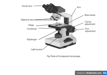
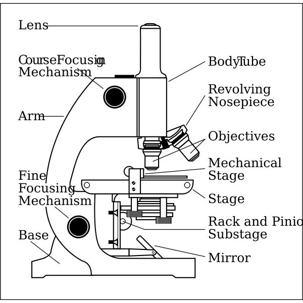
Parts of a microscope with functions and labeled diagram
Direct Antiglobulin Test | New Health Advisor Dispense one drop of RBC suspension (2%-5%) separately into each of the 4 test tubes, and label the 4 tubes with tags: Poly, Control, anti-IgG, and anti-C3. Wash the RBCs with saline, and then tilt the tube upside down to decant the saline. Then clear the bottoms of the tubes with the left saline, and repeat this for 3 to 4 times.

Simple doodles, Microscope parts, Microscopic images
Home | Royal Institution The Royal Institution is an independent charity dedicated to connecting people with the world of science, inspiring them to think more deeply about science and its place in our lives.

Compound Microscope- Definition, Labeled Diagram, Principle ...
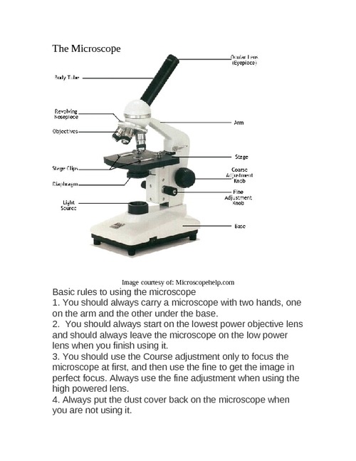
learning-center.homesciencetools.com › articleMicroscope Lab Experiments: An Introduction to the Microscope Microscope Worksheet: How to Record Microscope Observations In the field of science, recording observations while performing an experiment is one of the most useful tools available. Early scientists often kept very detailed journals of the experiments they performed, making entries for each individual experiment and writing down virtually ...

Label the microscope — Science Learning Hub
Motility Test (Theory) - Amrita Vishwa Vidyapeetham Virtual Lab In addition to being able to determine the presence or absence of motility, this method is useful in determining cellular shape (rod, coccus, or spiral) and arrangement (irregular clusters, packets, pairs, or long chains). 2. Hanging Drop slide

label the parts of the compound microscope - Brainly.ph
Ortur Laser Master 2 Pro Review - Is It Worth The Premium Price Tag? Let's do some further testing with the digital microscope. Here are the pictures taken with an Andonstar AD407 digital microscope . First thing that we see is that the Ortur Laser Master 2 Pro has a dot shape, sized a bit less than 0.1 times 0.2 millimetres.

Label the microscope — Science Learning Hub
Blood Cells & Formed Elements of the Blood | GetBodySmart Learn to identify cells under the microscope with these histology quizzes and labelling exercises. A monocyte in a section of capillary. The smalled formed elements are called platelets ( thrombocytes ). These are actually cytoplasmic fragments that pinch from large cells called megakaryocytes.

Compound Microscope Parts – Labeled Diagram and their ...
Biology Schemes of work term 1-3 Form 4 - Educationnewshub.co.ke Draw and label a transverse section of herbaceous and woody stems. Examine transverse section of a dicotyledonous stem, herbaceous and woody stems. Herbaceous stem, microscope, slides, Razors. KLB BK IV. PP 111-5 : 5: Stem tissues. Identify some stem tissues. Explain the role of stem tissues. Drawing and labeling diagrams; Discussion ...
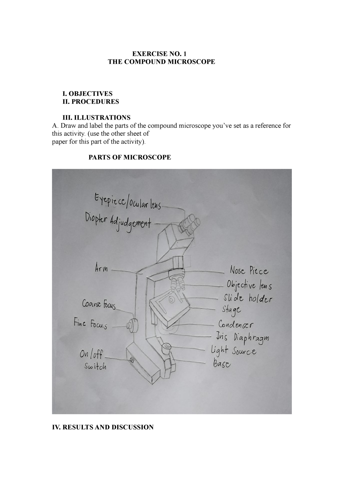
Compound microscopic microbiology - EXERCISE NO. 1 THE ...
rockyourhomeschool.net › microscope-worksheetsFree Microscope Worksheets for Simple Science Fun for Your ... Microscope Lab Report (drawing and notebooking page) You may print as many copies of these microscope worksheets that you’ll need for science fun with your kids, class, co-op, or community event. If you have a friend or co-worker who may like to use these free printable activities with their kids, I ask that you please share the link to this ...

Microscope Drawing Worksheet | Clipart library - Free Clipart ...
Wavefunction - BrainMass Wavefunction. A wave function describes a quantum state of particles and how they behave. The laws of quantum mechanics describe how the wave function changes over time. The common system for a wave function is Ѱ. Although Ѱ is a complex number, |Ѱ|² is a real number and corresponds to the probability density of finding a particle in a ...

Microscope Coloring | Clipart Panda - Free Clipart Images
Centromere - Genome.gov The centromere appears as a constricted region of a chromosome and plays a key role in helping the cell divide up its DNA during division (mitosis and meiosis). Specifically, it is the region where the cell's spindle fibers attach. Following attachment of the spindle fibers to the centromere, the two identical sister chromatids that make up ...
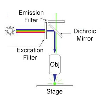
Fluorescence Microscopy - Explanation and Labelled Images ...
scheme work biology - Free KCPE Past Papers Introduction to light microscope. By the end of the lesson, the learner should be able to: Define a cell; Draw and label the light microscope; Description of a cell; Drawing and labeling the light microscope . Light microscope; Diagram of light microscope; Comprehensive secondary Biology students Bk. 1 page 17; Teachers bk. 1 pages 11-19; KLB ...

Label Microscope Diagram - EnchantedLearning.com
I'm having some issues with the crts on my electron microscope, I was ... Thank you, I was entirely at a loss before. There are a group of coaxial cables coming out of the machine. They seem to have been soldered into the circuitry by a third party. They're labeled as followed: Group 1 cables: B Input Xa Input Ya Input B Output Xa Output Ya Output 2 lines without labels . Group 2 cables Video In Video Out SEI Signal

Compound Microscope Parts – Labeled Diagram and their ...
Neuromuscular junction: Parts, structure and steps | Kenhub Neuromuscular junction (Synapsis neuromuscularis) At its simplest, the neuromuscular junction is a type of synapse where neuronal signals from the brain or spinal cord interact with skeletal muscle fibers, causing them to contract. The activation of many muscle fibers together causes muscles to contract, which in turn can produce movement.

Microscope With Labels Clip Art at Clker.com - vector clip ...
Cell Microscope Under Labeled Leaf [MUVB9S] draw and label a typical bacterial cell, then provide functions for at least five of the labeled structures add a drop of water (hypotonic solution) and a coverslip and observe the chloroplasts (green structures) and the cell walls harry potter fanfiction harry soul song students label the parts of a compound microscope the students will use a …

Microscope Diagram and Quiz | Science diagrams, Science ...
ECLIPSE Ti2 Series | Inverted Microscopes | Nikon Microscope Products ... The ECLIPSE Ti2 inverted microscope delivers an unparalleled 25mm field of view (FOV) that revolutionizes the way you see. With this incredible FOV, the Ti2 maximizes the sensor area of large-format CMOS cameras without making compromises, and significantly improves data throughput.

Compound Microscope Parts, Functions, and Labeled Diagram ...
Spiny chondrichthyan from the lower Silurian of South China Circles opposite the taxon labels show the presence of these characters at terminal nodes. ... (V27437.3 and V27437.5-7) were imaged with a GXM XTL3101 stereo microscope at the School of ...
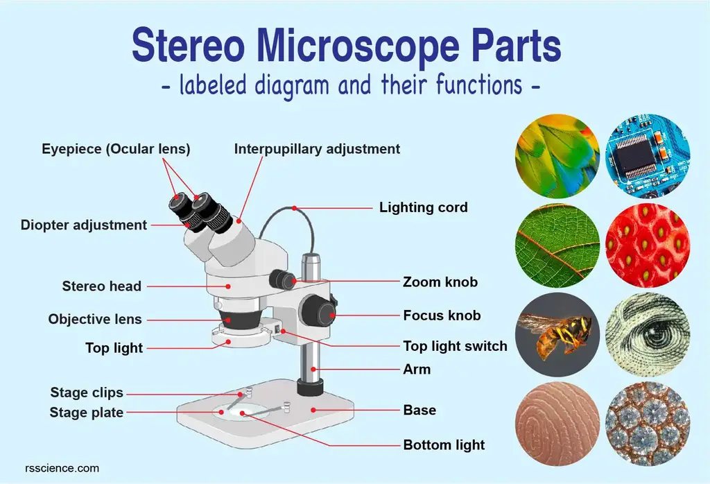
Parts of Stereo Microscope (Dissecting microscope) – labeled ...
Compound Microscope Review - ppt download
Microscope with labels picture

Simple Microscope - Diagram (Parts labelled), Principle ...

How to Draw a Microscope Easy | Sketches easy, Easy drawings ...
Answered: Microscope Structure and Function… | bartleby
Parts of a Microscope with Their Functions – Microbe Online

Parts of the Microscope with Labeling (also Free Printouts ...
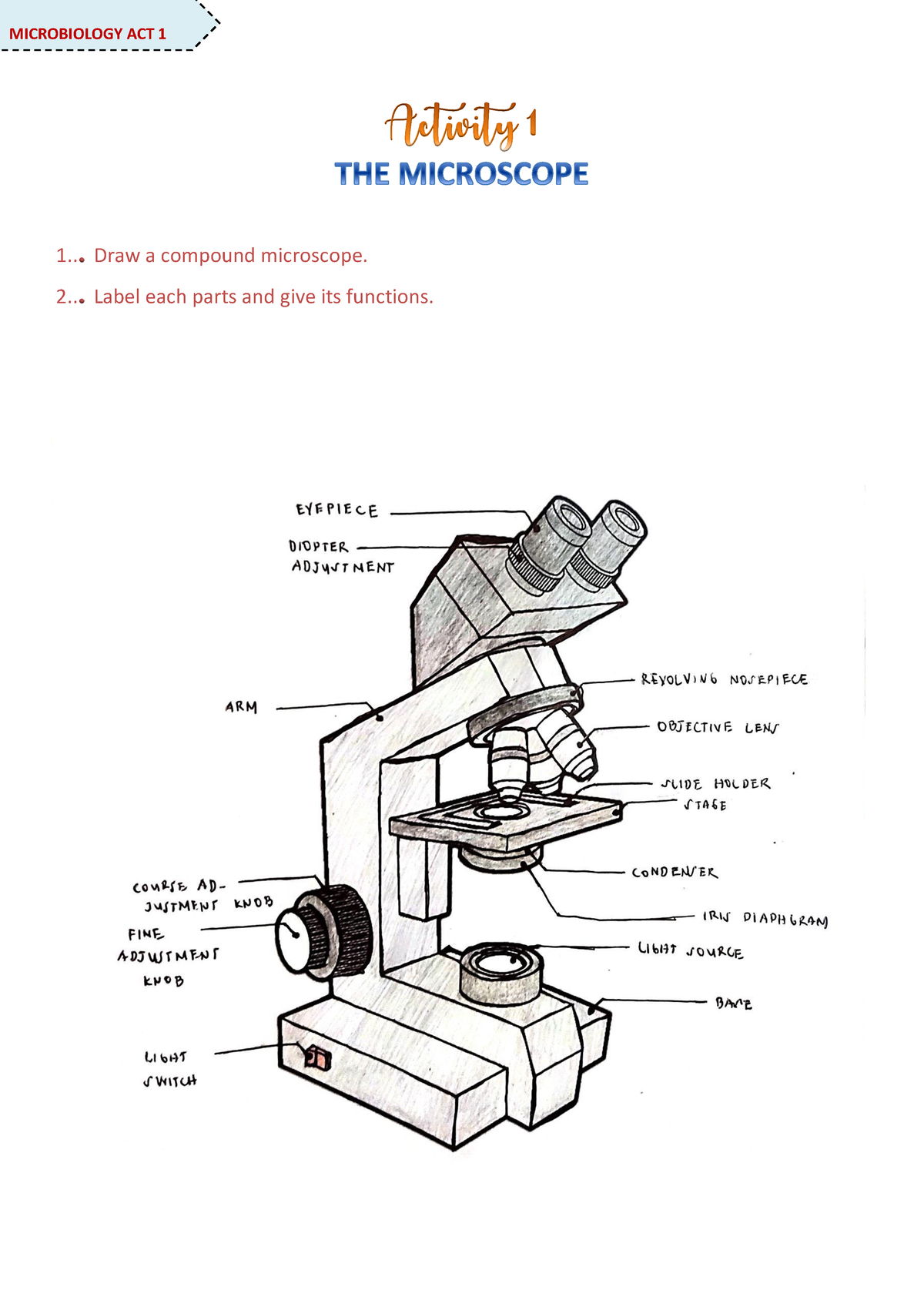
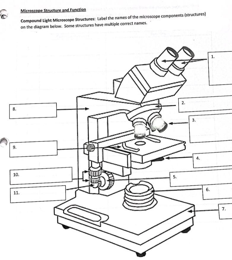
Microscope Activity - MICROBIOLOGY - 1... Draw a compound ...
Collection Of Free Microscopes Drawing Label Clipart ...
Microscope side vector drawing with parts labelled | Free SVG

How to draw compound of Microscope easily - step by step

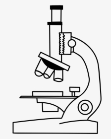
Free Microscope Drawing, Download Free Microscope Drawing png ...

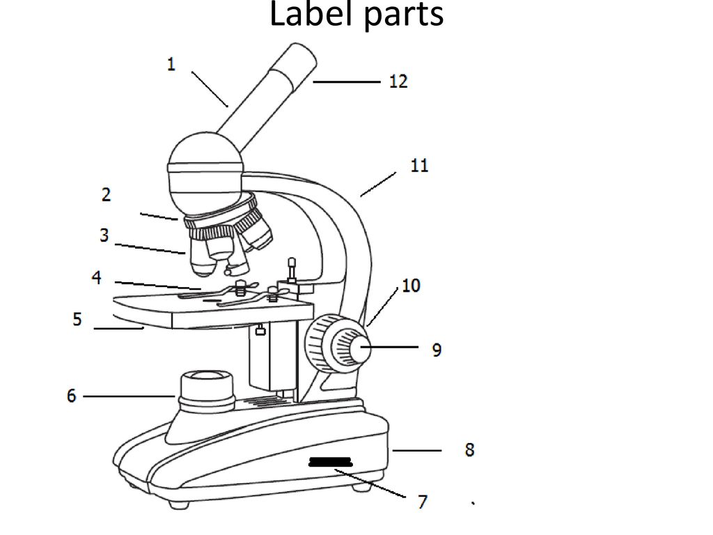
Microscope - Label - Part 2 Diagram | Quizlet
Parts of a microscope with functions and labeled diagram
Free Microscope Drawing, Download Free Microscope Drawing png ...
Free Microscope Drawing, Download Free Microscope Drawing png ...

Microscope Parts Review Diagram | Quizlet

Microscope Diagram Labeled, Unlabeled and Blank | Parts of a ...

MICROSCOPE Labeling - Part - 3

Microscope Labeling

Free Microscope Drawing, Download Free Microscope Drawing png ...

Label the Parts of the Microscope - Brainly.ph

Compound Microscope Parts – Labeled Diagram and their ...

A Study of the Microscope and its Functions With a Labeled ...
compound-microscope-unlabeled.jpg

Draw the diagram of a microscope and label its parts.

Dissecting Stereo Microscope Parts and Functions

Parts of a microscope with functions and labeled diagram
Post a Comment for "45 microscope drawing and label"