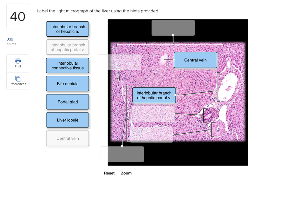
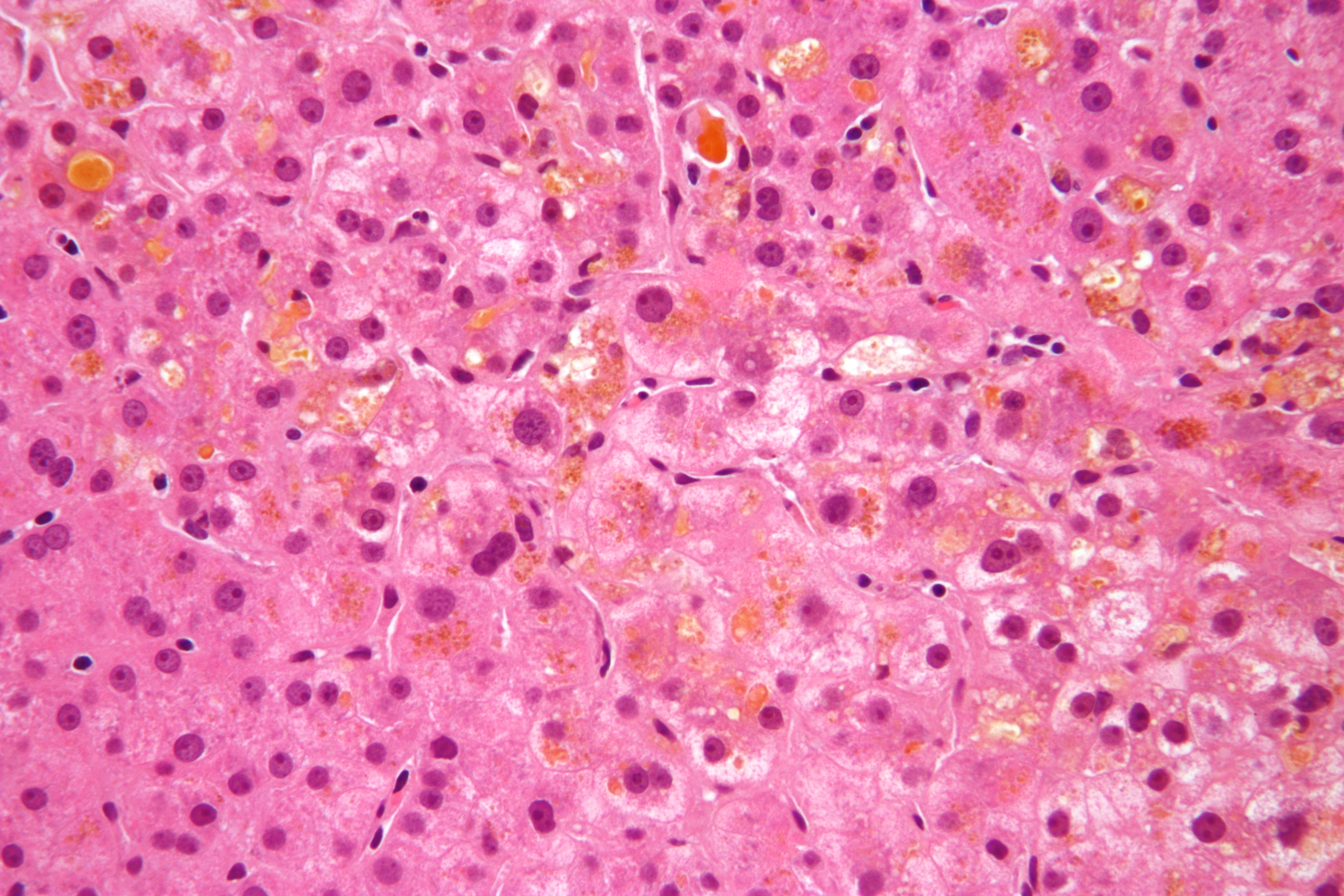
40 label the light micrograph of the liver using the hints provided
Label the light micrograph of the seminiferous tubule using ... Start studying Label the light micrograph of the seminiferous tubule using the hints provided. Learn vocabulary, terms, and more with flashcards, games, and other study tools. Solved Label the light micrograph of the liver using the ... Expert Answer. 100% (5 ratings) Transcribed image text: Label the light micrograph of the liver using the hints provided. Interlobular branch of hepatic a 0.19 points Interlobular branch of hepatic portal v Central vein Interlobular connective tissue Print References Bile ductule Interlobular branch of hepatic portal v.
Solved Label the light micrograph of the colon using the ... Expert Answer. 94% (18 ratings) From above downward: Openin …. View the full answer. Transcribed image text: Label the light micrograph of the colon using the hints provided Muscularis externa Intestinal gland Muscularis mucosae Goblet cell Submucosa Mucosa Opening df intestinal land Reset Zoom. Previous question Next question.

Label the light micrograph of the liver using the hints provided
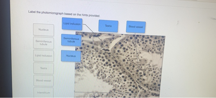
Label the Stomach and Duodenum Using the Hints if Provided. Apr 19, 2022 · Label the structures of the posterior thoracic wall using the hints if provided. Body Fundus Cardiac Pylorus Esophagus Stomach Reset Zoom Prav 1 of 10 C J Jone. Testis Layer 2 Label the testis and spermatic cord using the hints. Label the light micrograph of the colon using the hints provided. Label the internal features of stomach and duodenum ... Digestive lab Flashcards | Quizlet Label the mucous membrane tissue from the stomach using the hints if provided. Label the digestive abdominal contents using the hints if provided. Place the appropriate words and descriptions with the picture with the correct highlighted digestive accessory organ. Label the structures of the posterior thoracic wall using the hints if provided. Label The Photomicrograph Based On The Hints Provided ... Free answer to label the structures in the photomicrograph based on the hints provided. Label the photomicrograph based on the hints provided. Label the photomicrograph based on the hints provided. Distinguish the different types of pituitary cells using the light microscope and electron microscope. Spleen capsule capsule white pulp.
Label the light micrograph of the liver using the hints provided. Label The Photomicrograph Based On The Hints Provided ... Free answer to label the structures in the photomicrograph based on the hints provided. Label the photomicrograph based on the hints provided. Label the photomicrograph based on the hints provided. Distinguish the different types of pituitary cells using the light microscope and electron microscope. Spleen capsule capsule white pulp. Digestive lab Flashcards | Quizlet Label the mucous membrane tissue from the stomach using the hints if provided. Label the digestive abdominal contents using the hints if provided. Place the appropriate words and descriptions with the picture with the correct highlighted digestive accessory organ. Label the structures of the posterior thoracic wall using the hints if provided. Label the Stomach and Duodenum Using the Hints if Provided. Apr 19, 2022 · Label the structures of the posterior thoracic wall using the hints if provided. Body Fundus Cardiac Pylorus Esophagus Stomach Reset Zoom Prav 1 of 10 C J Jone. Testis Layer 2 Label the testis and spermatic cord using the hints. Label the light micrograph of the colon using the hints provided. Label the internal features of stomach and duodenum ...
Eawag - Swiss Federal Institute of Aquatic Science and ...

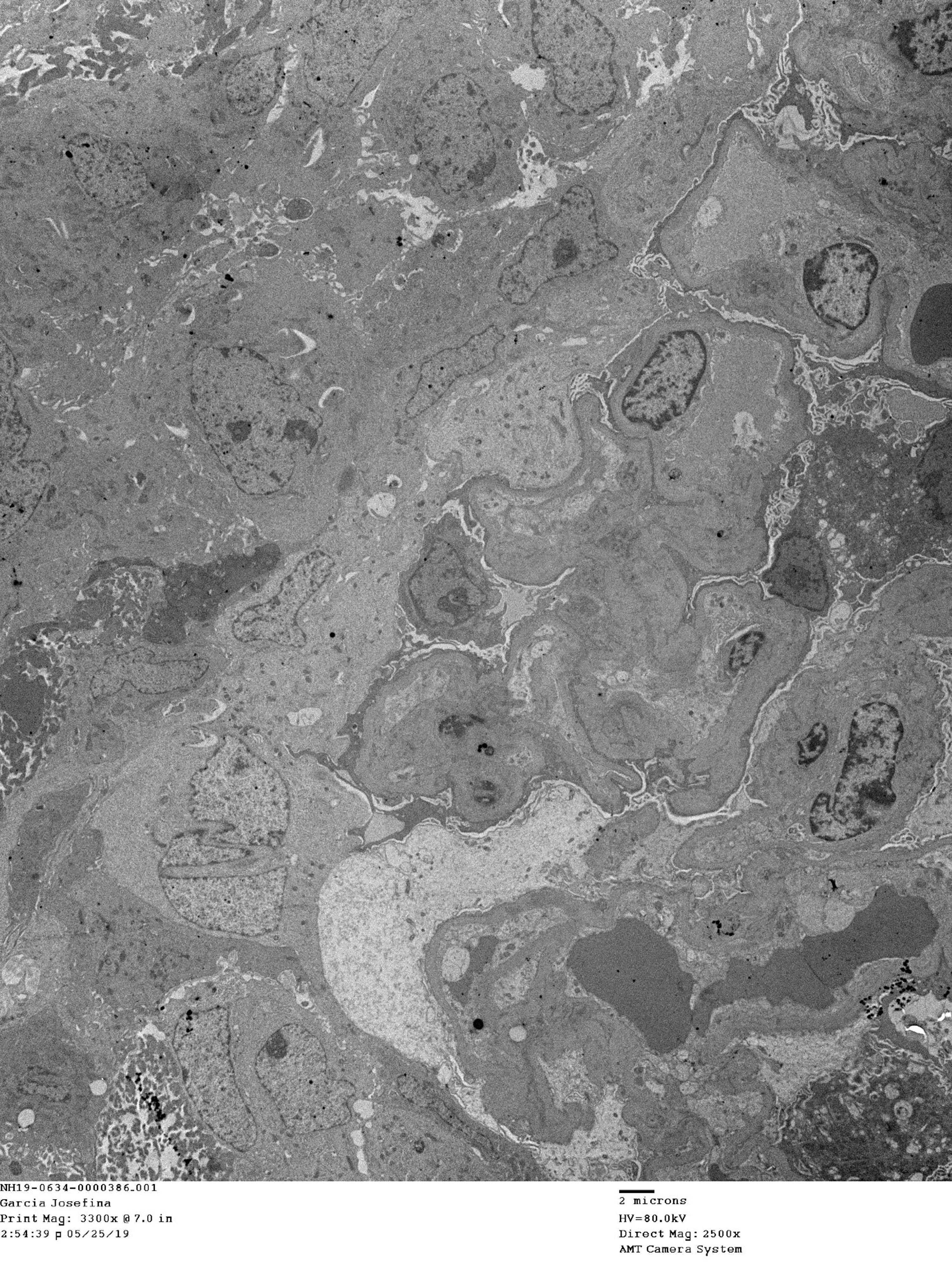
Ion channels and channelopathies in glomeruli | Physiological ...
Solved Label the photomicrograph based on the hints | Chegg.com
Prussian blue technique is prone to yield false negative ...

A&P 2 Lab Practical Final Flashcards | Quizlet
Cerebral Corpora amylacea are dense membranous labyrinths ...

Endometriosis genetic discovery may lead to new forms of ...

Digestive lab Flashcards | Quizlet

Topic 1.3 Membrane Structure - AMAZING WORLD OF SCIENCE WITH ...
Release of Hepatic Plasmodium yoelii Merozoites into the ...
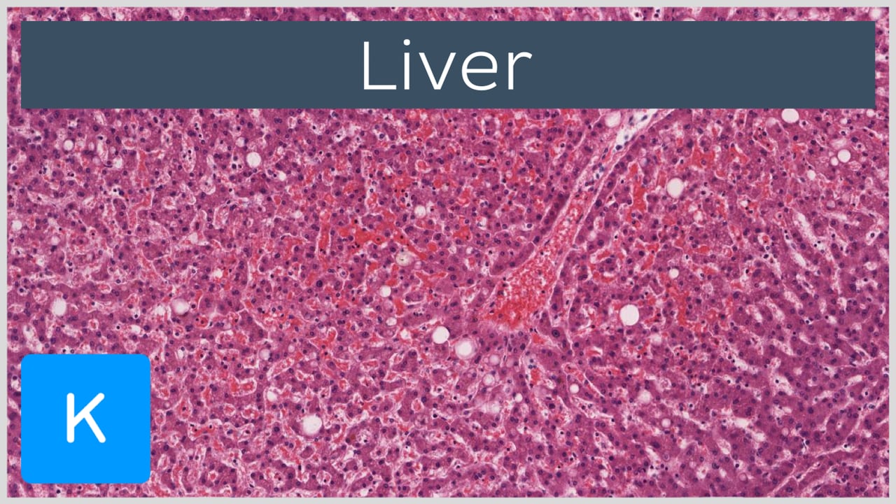
Liver histology
Development of biotechnology applied to food and health, to ...
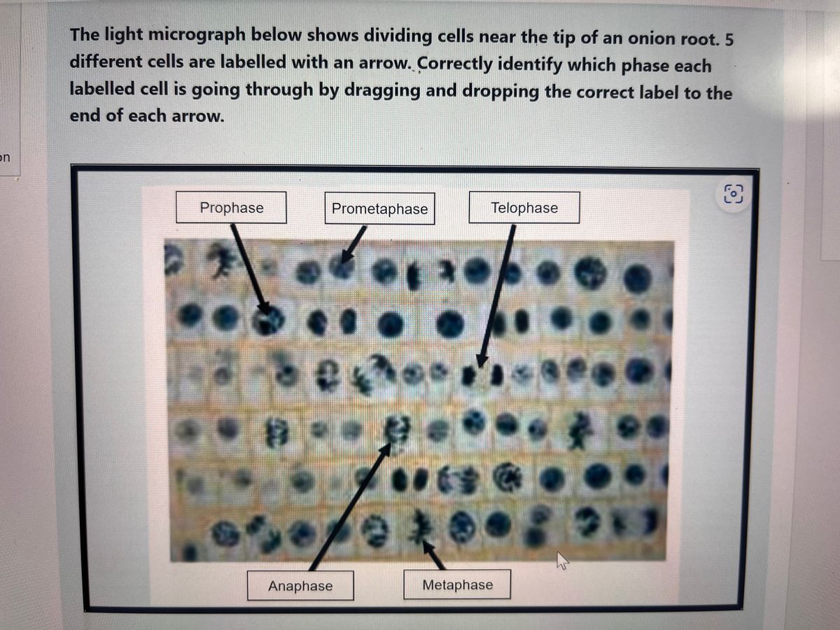
Answered: Correctly identify which phase each | bartleby

Epoxy Resin Embedding of Animal and Human Tissues for ...

Nanoscale magnetic imaging of ferritin in a single cell

Spatially resolved cell polarity proteomics of a human ...
Relative expression of microRNAs, apoptosis, and ...
Solved Label the light micrograph of the liver using the ...

ASAL Biology exec preview_Digital by Cambridge University ...
Rapid detection of hysteromyoma and cervical cancer based ...

Photomicrographs - an overview | ScienceDirect Topics

GI Post Lab Flashcards | Quizlet

Digestive lab Flashcards | Quizlet

Hunting coronavirus by transmission electron microscopy – a ...
Strange Infatuation - Perfect Abstraction | OpenSea

Upper panels: a representative light micrograph of a resected ...
Sharing scientific knowledge through publications: what do ...
Cholestasis - Wikipedia

Spatial Transcriptomics-correlated Electron Microscopy | bioRxiv
Full article: In vitro assays and biomarkers for drug-induced ...
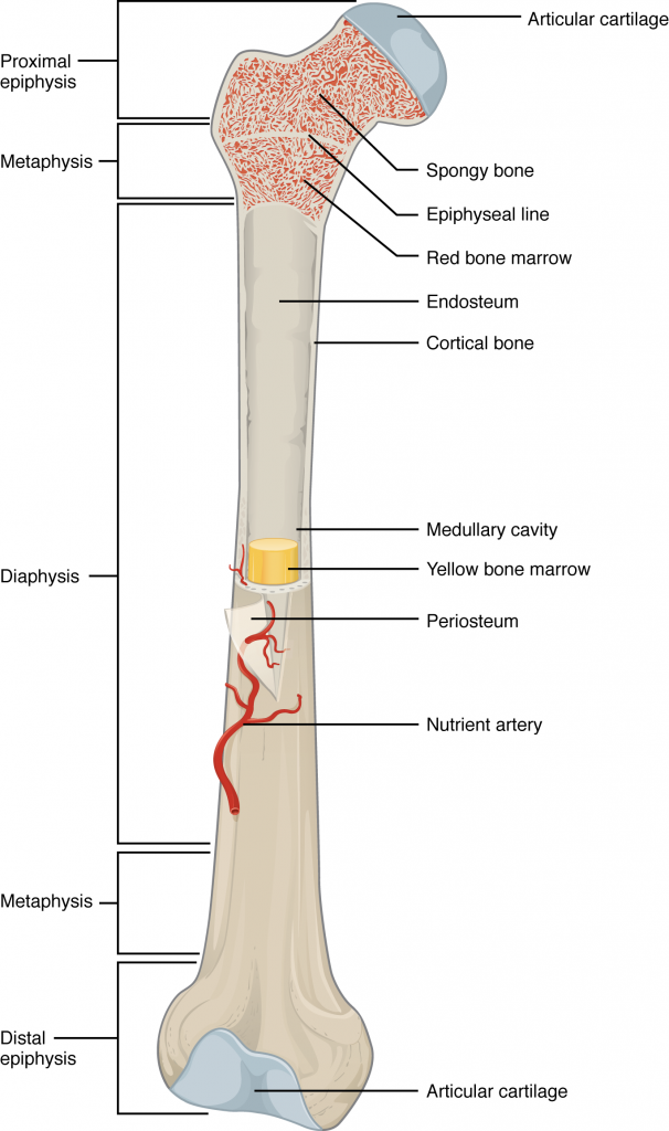
6.3 Bone Structure – Anatomy & Physiology

Multiple Myeloma: Causes, First Signs, Symptoms, Diagnosis ...

30 Cellular Respiration Photos and Premium High Res Pictures ...

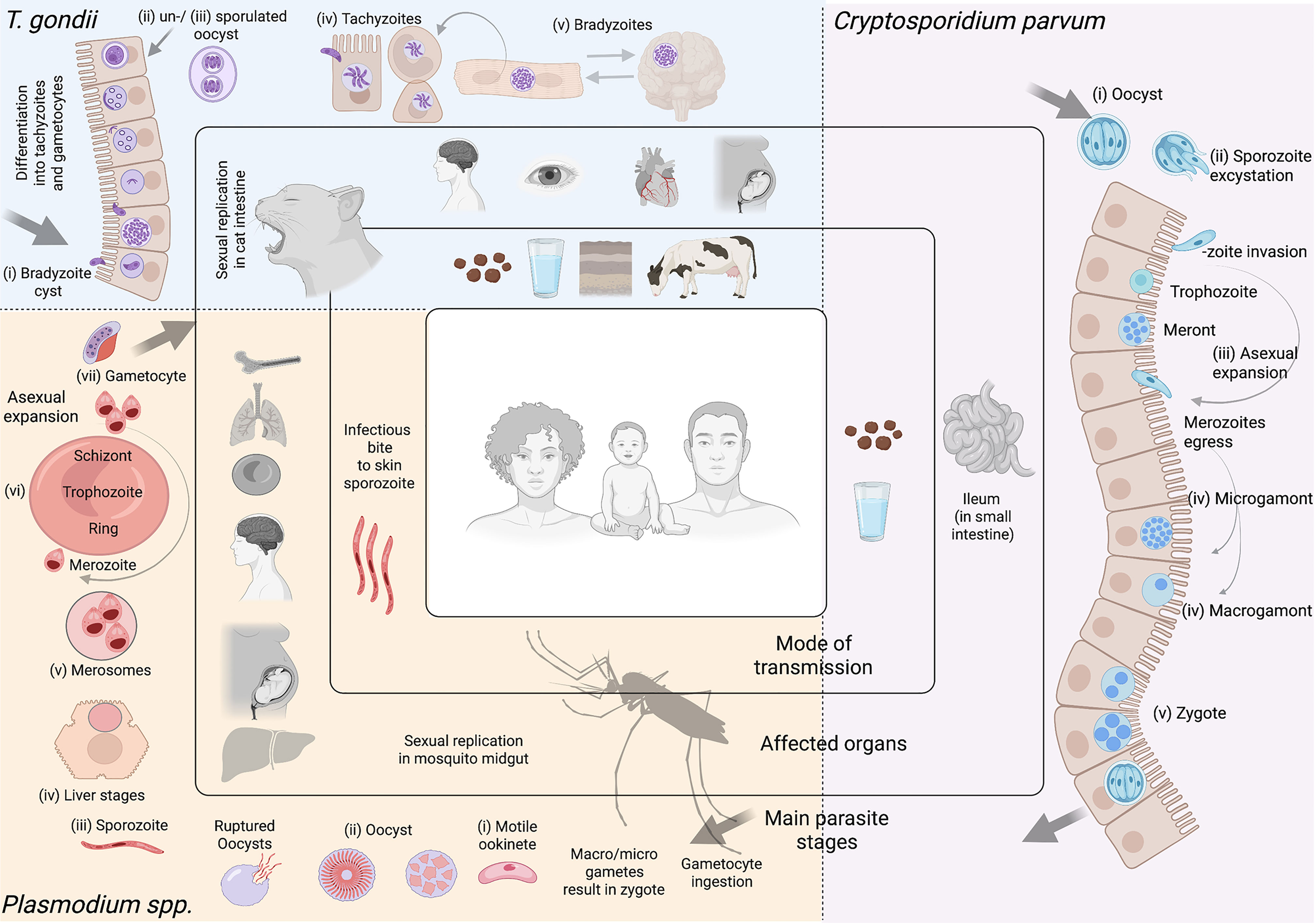
High resolution 3D perspective of Plasmodium biology ...
Cureus | Tofacitinib-Induced Antineutrophil Cytoplasmic ...
Frontiers | Paving the Way: Contributions of Big Data to ...

Light Micrograph of Lung Cancer Cells' Photographic Print ...

Correlative light and electron microscopy methods for the ...
Welcome To Netter Images

Digestive System Flashcards | Quizlet
Post a Comment for "40 label the light micrograph of the liver using the hints provided"