45 label the transmission electron micrograph
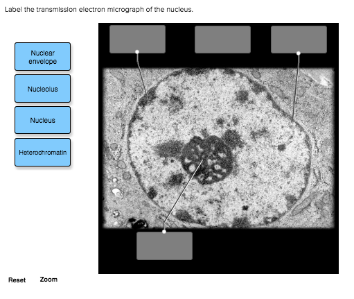
Labeling the Cell Flashcards | Quizlet Image: Label the transmission electron micrograph of the nucleus. membrane bound organelles. golgi apparatus, mitochondrion ... Transmission electron microscopy - Wikipedia Transmission electron microscopy (TEM) is a microscopy technique in which a beam of electrons is transmitted through a specimen to form an image.
Solved Label the transmission electron micrograph of the | Chegg.com Question: Label the transmission electron micrograph of the mitochondrion. Matrix granule Mitochondrion Outer membrane Cristae Inner membrane Matrix Reset Zoom.
Label the transmission electron micrograph
The Transmission Electron Microscope | CCBER Transmission electron microscopes (TEM) are microscopes that use a particle beam of electrons to visualize specimens and generate a highly-magnified image. TEMs ... TRANSMISSION ELECTRON MICROSCOPE - TRI-Genotoul A high-energy electron beam is transmitted through a thin sample and the image is formed on a phosphorescent screen coupled to a digital camera. TEM has many ... Label the transmission electron micrograph of the cell. 0 ... - Chegg Question: Label the transmission electron micrograph of the cell. 0 Nucleus rences Mitochondrion Heterochromatin Peroxisome Vesicle ULAR bumit Click and ...
Label the transmission electron micrograph. anatomy 10.png - Label the transmission electron micrograph of the View anatomy 10.png from ZOOL 1090 at Utah Valley University. Label the transmission electron micrograph of the. 8.2: Transmission Electron Microscopy - Chemistry LibreTexts Aug 28, 2022 ... Transmission electron microscopy (TEM) is a form of microscopy which in which a beam of electrons transmits through an extremely thin ... (A and B) Electron micrograph of a cell labeled for/5-tubulin followed... No microtubule labeling is evident. ... Fluorescence microscopy in combination with TEM and an ion beam analysis (IBA, which allows the evaluation of the ... Site-specific labeling of proteins for electron microscopy - PMC - NCBI Sep 25, 2015 ... The eluted conjugate is now ready for visualization by negative stain or cryo electron microscopy. (D) A representative micrograph containing ...
Label the transmission electron micrograph of the cell. 0 ... - Chegg Question: Label the transmission electron micrograph of the cell. 0 Nucleus rences Mitochondrion Heterochromatin Peroxisome Vesicle ULAR bumit Click and ... TRANSMISSION ELECTRON MICROSCOPE - TRI-Genotoul A high-energy electron beam is transmitted through a thin sample and the image is formed on a phosphorescent screen coupled to a digital camera. TEM has many ... The Transmission Electron Microscope | CCBER Transmission electron microscopes (TEM) are microscopes that use a particle beam of electrons to visualize specimens and generate a highly-magnified image. TEMs ...
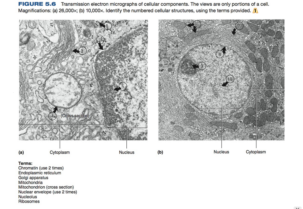
Solved FIGURE 5.6 Transmission electron micrographs of ...
Scanning Transmission Electron Micrograph (STEM). Vesicles ...
Click-correlative light and electron microscopy (click-AT ...

Transmission electron micrograph of mature MRCs with anti-Na ...

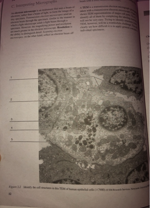
lab 12 word.docx - Name: Laboratory Assessment Date: 12 ...
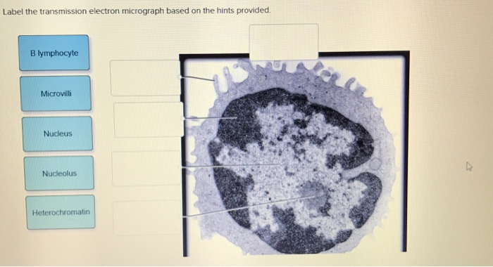
Solved Label the transmission electron micrograph based on ...

Transmission electron micrograph of an animal cell - Stock ...
Electron Microscopy – Centro Investigación Príncipe Felipe

TEM of animal cell - Stock Image - G450/0055 - Science Photo ...
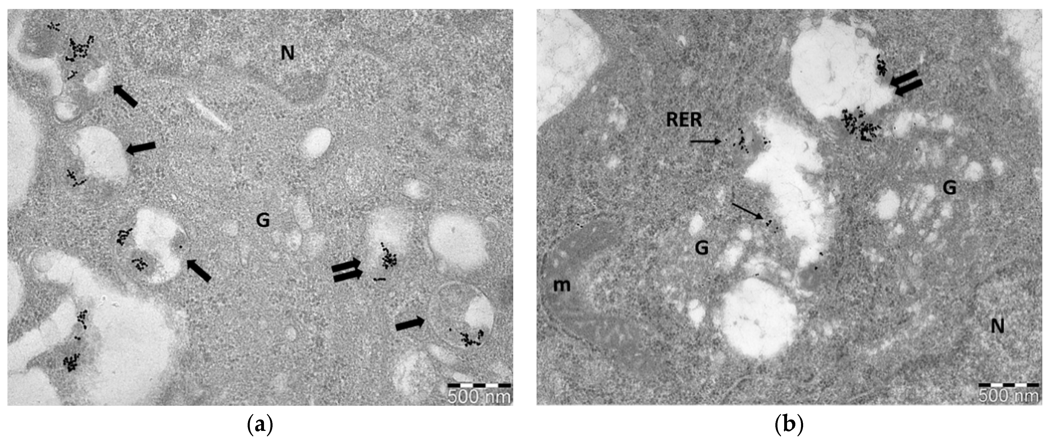
Biology | Free Full-Text | Immuno-Electron and Confocal Laser ...

Solved Label the transmission electron micrograph based on ...
BIOL 230 Lecture Guide - Electron Micrograph of Rough ...

Transmission electron micrograph of turkey spermatozoa ...

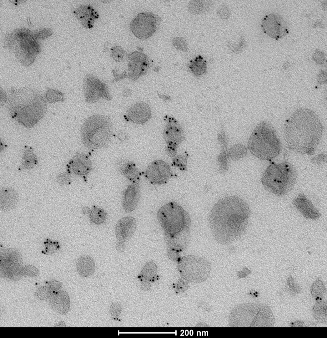
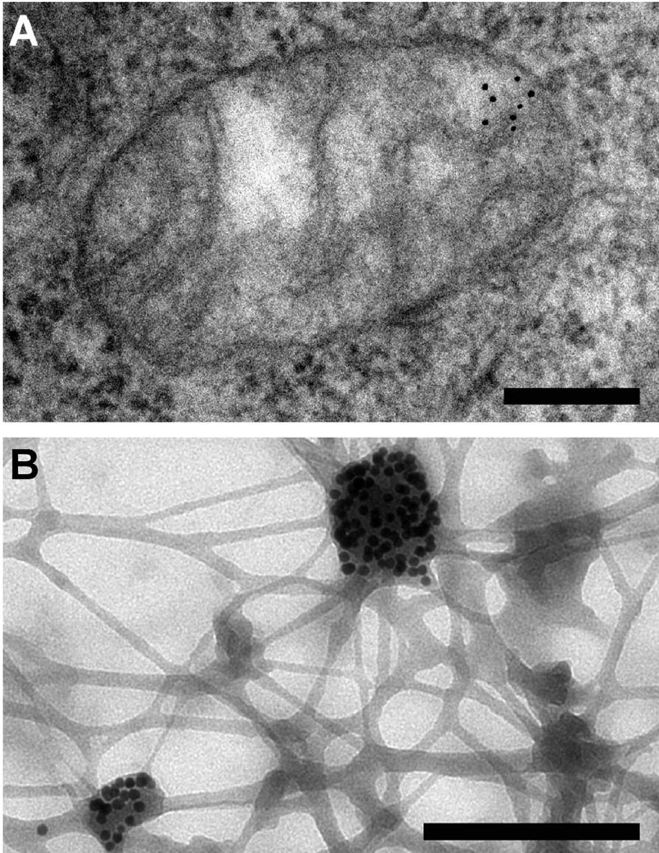
Electron micrographs of negative staining and immunogold ...
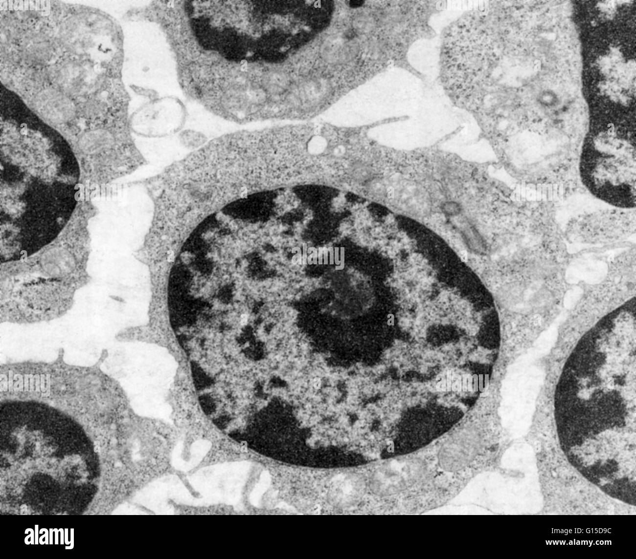
Nucleus and nucleolus, TEM stock photo. Image of cytology ...
Transmission electron microscope (TEM) micrograph showing a ...
Solved A TEME is a transmission electron micro taken with a ...

A&P Unit 2 Exam Flashcards | Quizlet
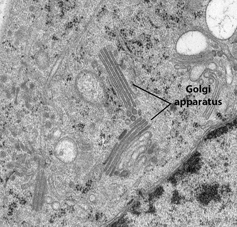
BIOL 230 Lecture Guide - Electron Micrograph of a Golgi Body

Figure, Transmission Electron Micrograph of Rough Endoplasmic ...
Nanomaterials | Free Full-Text | A Guide for Using ...
Solved Label the transmission electron micrograph of the ...
Transmission electron microscopy Black and White Stock Photos ...
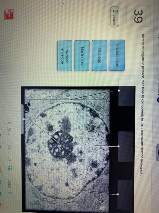
Solved Identify the organelle pictured, then label its ...

Labeling the Cell Flashcards | Quizlet

Labeling the Cell Flashcards | Quizlet

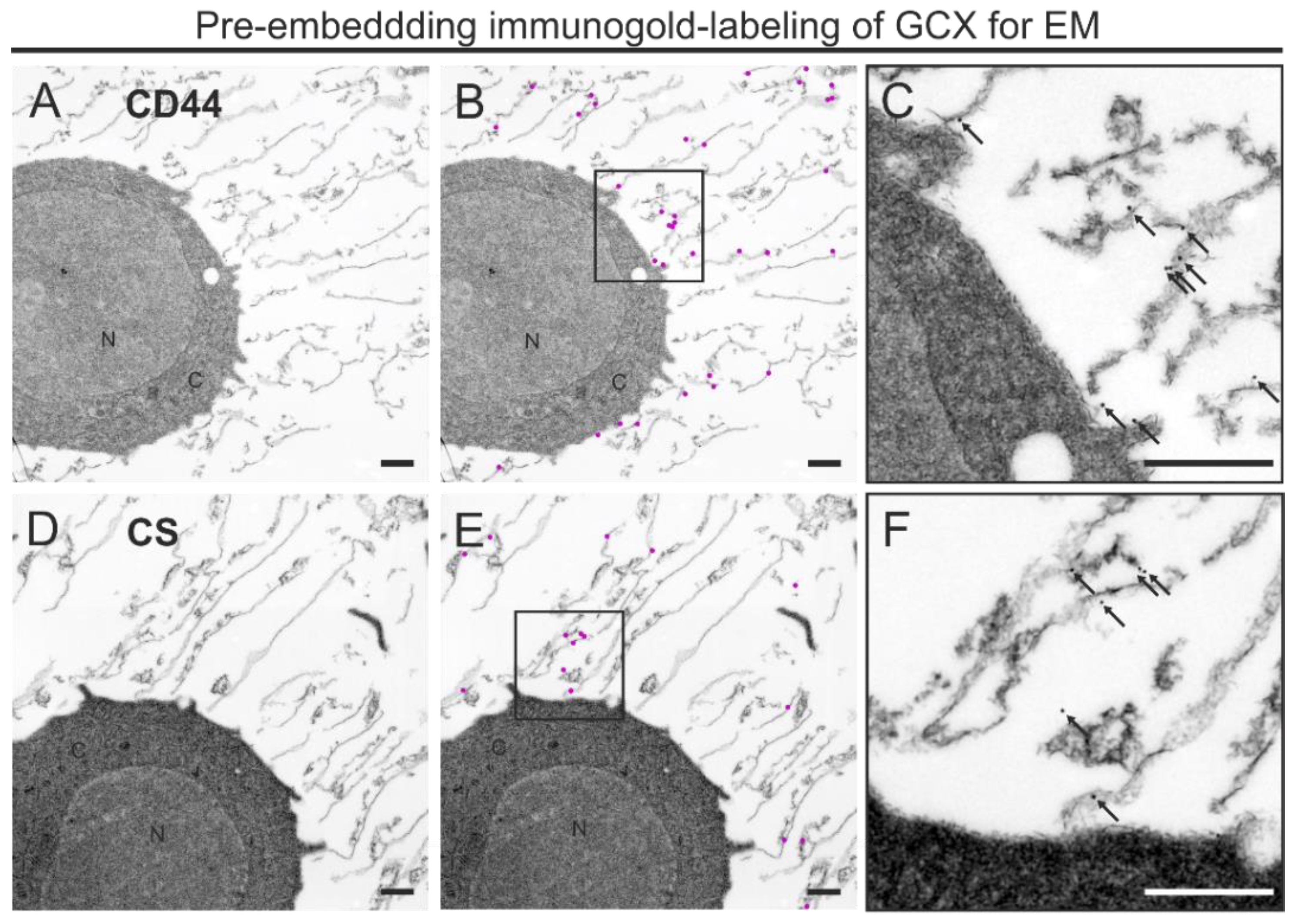
Transmission electron microscopy images of immunogold-labeled ...

Transmission electron micrograph of endocytosed gold-ASOR ...
Immunogold labelling - Wikipedia

The transmission electronmicroscope shows parts of two ...

The Transmission Electron Microscope | CCBER

Pinocytosis. endothelial cell. Transmission electron ...

Immune electron microscopy - Wikipedia

Biology 2e, The Cell, Cell Structure, Eukaryotic Cells ...

Labeling the Cell Flashcards | Quizlet
Microscopy Innovations | Transmission electron microscopy (TEM)

Transmission electron micrograph of cells infected with ...
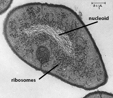
BIOL 230 Lecture Guide - Prokaryotic Ribosome

Transmission electron microscopy (TEM) imaging of nuclear ...

Labeling the Cell Flashcards | Quizlet

Muscle Lab 19 Figure 19.5 Sarcomere Diagram | Quizlet

Transmission electron micrograph of bladder cancer exosomes ...

BSC2085L Lab 8 Exercises 12, 13, & 14 Flashcards | Quizlet

Hunting coronavirus by transmission electron microscopy – a ...

Solved Mitochondrion Nucleus Vesicle Peroxisome | Chegg.com
Post a Comment for "45 label the transmission electron micrograph"